


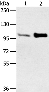
Figure 1. Western blot analysis of ACTN2 using anti-ACTN2 antibody (A03673-1). Electrophoresis was performed on a 5-20% SDS-PAGE gel at 70V (Stacking gel) / 90V (Resolving gel) for 2-3 hours. The sample well of each lane was loaded with 30 ug of sample under reducing conditions. Lane 1: human Hela whole cell lysates, Lane 2: human HEPG2 whole cell lysates, Lane 3: rat skeletal muscle tissue lysates, Lane 4: rat heart tissue lysates, Lane 5: mouse skeletal muscle tissue lysates, Lane 6: mouse heart tissue lysates. After electrophoresis, proteins were transferred to a nitrocellulose membrane at 150 mA for 50-90 minutes. Blocked the membrane with 5% non-fat milk/TBS for 1.5 hour at RT. The membrane was incubated with rabbit anti-ACTN2 antigen affinity purified polyclonal antibody (Catalog # A03673-1) at 0.5 microg/mL overnight at 4°C, then washed with TBS-0.1%Tween 3 times with 5 minutes each and probed with a goat anti-rabbit IgG-HRP secondary antibody at a dilution of 1:5000 for 1.5 hour at RT. The signal is developed using an Enhanced Chemiluminescent detection (ECL) kit (Catalog # EK1002) with Tanon 5200 system. A specific band was detected for ACTN2 at approximately 104 kDa. The expected band size for ACTN2 is at 104 kDa.
. ACTN2 was detected in a paraffin-embedded section of mouse cardiac tissue. Heat mediated antigen retrieval was performed in EDTA buffer (pH 8.0, epitope retrieval solution). The tissue section was blocked with 10% goat serum. The tissue section was then incubated with 2 microg/ml rabbit anti-ACTN2 Antibody (A03673-1) overnight at 4°C. Biotinylated goat anti-rabbit IgG was used as secondary antibody and incubated for 30 minutes at 37°C. The tissue section was developed using Strepavidin-Biotin-Complex (SABC) (Catalog # SA1022) with DAB as the chromogen.")
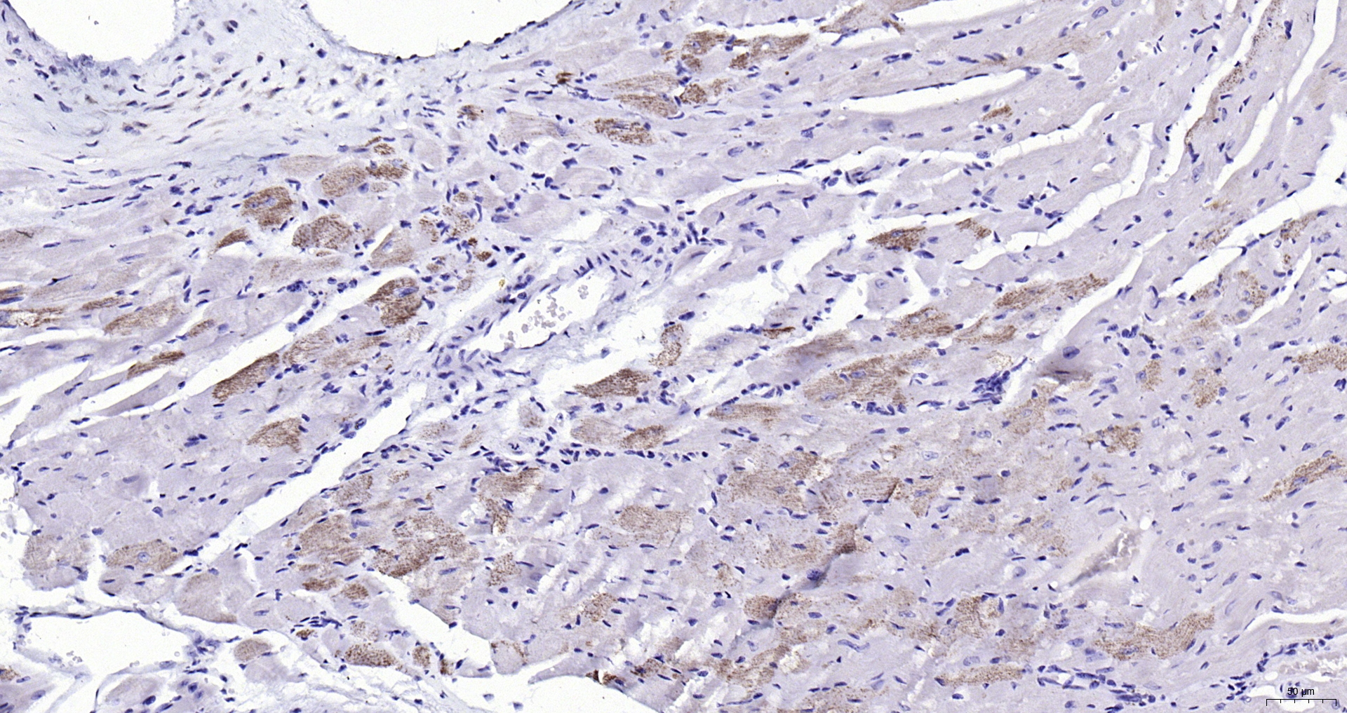
. ACTN2 was detected in a paraffin-embedded section of rat cardiac tissue. Heat mediated antigen retrieval was performed in EDTA buffer (pH 8.0, epitope retrieval solution). The tissue section was blocked with 10% goat serum. The tissue section was then incubated with 2 microg/ml rabbit anti-ACTN2 Antibody (A03673-1) overnight at 4°C. Biotinylated goat anti-rabbit IgG was used as secondary antibody and incubated for 30 minutes at 37°C. The tissue section was developed using Strepavidin-Biotin-Complex (SABC) (Catalog # SA1022) with DAB as the chromogen.")
. Overlay histogram showing A431 cells stained with A03673-1 (Blue line). To facilitate intracellular staining, cells were fixed with 4% paraformaldehyde and permeabilized with permeabilization buffer. The cells were blocked with 10% normal goat serum. And then incubated with rabbit anti-ACTN2 Antibody (A03673-1, 1 microg/1x106 cells) for 30 min at 20°C. DyLight®488 conjugated goat anti-rabbit IgG (BA1127, 5-10 microg/1x106 cells) was used as secondary antibody for 30 minutes at 20°C. Isotype control antibody (Green line) was rabbit IgG (1 microg/1x106) used under the same conditions. Unlabelled sample without incubation with primary antibody and secondary antibody (Red line) was used as a blank control.")
. Overlay histogram showing ANA-1 cells stained with A03673-1 (Blue line). To facilitate intracellular staining, cells were fixed with 4% paraformaldehyde and permeabilized with permeabilization buffer. The cells were blocked with 10% normal goat serum. And then incubated with rabbit anti-ACTN2 Antibody (A03673-1, 1 microg/1x106 cells) for 30 min at 20°C. DyLight®488 conjugated goat anti-rabbit IgG (BA1127, 5-10 microg/1x106 cells) was used as secondary antibody for 30 minutes at 20°C. Isotype control antibody (Green line) was rabbit IgG (1 microg/1x106) used under the same conditions. Unlabelled sample without incubation with primary antibody and secondary antibody (Red line) was used as a blank control.")
Figure 1. Western blot analysis of ACTN2 using anti-ACTN2 antibody (A03673-1). Electrophoresis was performed on a 5-20% SDS-PAGE gel at 70V (Stacking gel) / 90V (Resolving gel) for 2-3 hours. The sample well of each lane was loaded with 30 ug of sample under reducing conditions. Lane 1: human Hela whole cell lysates, Lane 2: human HEPG2 whole cell lysates, Lane 3: rat skeletal muscle tissue lysates, Lane 4: rat heart tissue lysates, Lane 5: mouse skeletal muscle tissue lysates, Lane 6: mouse heart tissue lysates. After electrophoresis, proteins were transferred to a nitrocellulose membrane at 150 mA for 50-90 minutes. Blocked the membrane with 5% non-fat milk/TBS for 1.5 hour at RT. The membrane was incubated with rabbit anti-ACTN2 antigen affinity purified polyclonal antibody (Catalog # A03673-1) at 0.5 microg/mL overnight at 4°C, then washed with TBS-0.1%Tween 3 times with 5 minutes each and probed with a goat anti-rabbit IgG-HRP secondary antibody at a dilution of 1:5000 for 1.5 hour at RT. The signal is developed using an Enhanced Chemiluminescent detection (ECL) kit (Catalog # EK1002) with Tanon 5200 system. A specific band was detected for ACTN2 at approximately 104 kDa. The expected band size for ACTN2 is at 104 kDa.
Anti-ACTN2 Antibody Picoband(r)
A03673-1-CARRIER-FREE
ApplicationsFlow Cytometry, Western Blot, ELISA, ImmunoHistoChemistry
Product group Antibodies
ReactivityHuman, Mouse, Rat
TargetACTN2
Overview
- SupplierBoster Bio
- Product NameAnti-ACTN2 Antibody Picoband(r)
- Delivery Days Customer9
- ApplicationsFlow Cytometry, Western Blot, ELISA, ImmunoHistoChemistry
- CertificationResearch Use Only
- ClonalityPolyclonal
- Concentration500 ug/ml
- Gene ID88
- Target nameACTN2
- Target descriptionactinin alpha 2
- Target synonymsCMD1AA, CMH23, CMYO8, CMYP8, MPD6, MYOCOZ, alpha-actinin-2, F-actin cross-linking protein, alpha-actinin skeletal muscle
- HostRabbit
- IsotypeIgG
- Protein IDP35609
- Protein NameAlpha-actinin-2
- Scientific DescriptionBoster Bio Anti-ACTN2 Antibody Picoband® catalog # A03673-1. Tested in ELISA, Flow Cytometry, IHC, WB applications. This antibody reacts with Human, Mouse, Rat. The brand Picoband indicates this is a premium antibody that guarantees superior quality, high affinity, and strong signals with minimal background in Western blot applications. Only our best-performing antibodies are designated as Picoband, ensuring unmatched performance.
- ReactivityHuman, Mouse, Rat
- Storage Instruction-20°C,2°C to 8°C
- UNSPSC12352203
Related products
Product group Antibodies
Anti-ACTN2 AntibodyA38104
ApplicationsWestern Blot, ImmunoHistoChemistry
ReactivityHuman, Mouse
- SizePrice
Product group Antibodies
Anti-ACTN2 Antibody144-03718
ApplicationsImmunoFluorescence, Western Blot, ImmunoHistoChemistry
ReactivityHuman, Mouse, Rat
TargetACTN2
- SizePrice
Product group Antibodies
References
ApplicationsImmunoFluorescence, Western Blot, ELISA, ImmunoCytoChemistry, ImmunoHistoChemistry, ImmunoHistoChemistry Frozen, ImmunoHistoChemistry Paraffin
ReactivityBovine, Canine, Chicken, Equine, Human, Mouse, Porcine, Rabbit, Rat, Sheep
TargetACTN2
- SizePrice
Product group Antibodies
ACTN2 AntibodyCSB-PA001242LA01HU
ApplicationsWestern Blot, ELISA, ImmunoHistoChemistry
ReactivityHuman, Rat
TargetACTN2
- SizePrice
Product group Antibodies
ACTN2 Polyclonal AntibodyCAC15868
ApplicationsWestern Blot, ELISA, ImmunoHistoChemistry
ReactivityRat
TargetACTN2
- SizePrice
Product group Antibodies

ACTN2 AntibodyLS-C403508
ApplicationsWestern Blot, ELISA, ImmunoHistoChemistry
ReactivityHuman, Mouse
TargetACTN2
- SizePrice
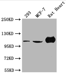
![Various whole cell extracts (30 μg) were separated by 5% SDS-PAGE, and the membrane was blotted with alpha Actinin 2 antibody [N1N3] (GTX103219) diluted at 1:1000. The HRP-conjugated anti-rabbit IgG antibody (GTX213110-01) was used to detect the primary antibody.](https://www.genetex.com/upload/website/prouct_img/normal/GTX103219/GTX103219_44335_20210702_WB_M_22072519_433.webp)
Product group Antibodies
alpha Actinin 2 antibody [N1N3]GTX103219
ApplicationsImmunoFluorescence, Western Blot, ImmunoCytoChemistry, ImmunoHistoChemistry, ImmunoHistoChemistry Paraffin
ReactivityHuman, Mouse, Porcine, Rat, Zebra Fish
TargetACTN2
- SizePrice

Product group Antibodies
Anti-ACTN2 AntibodyHPA008315
ApplicationsImmunoHistoChemistry
ReactivityHuman
TargetACTN2
- SizePrice
Product group Antibodies
TargetACTN2
- SizePrice