

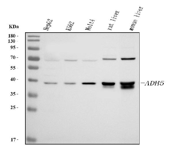
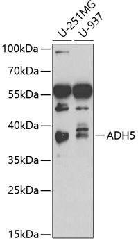
Figure 1. Western blot analysis of ADH5 using anti-ADH5 antibody (RP1111). Electrophoresis was performed on a 5-20% SDS-PAGE gel at 70V (Stacking gel) / 90V (Resolving gel) for 2-3 hours. The sample well of each lane was loaded with 30 ug of sample under reducing conditions. Lane 1: human HepG2 whole cell lysates, Lane 2: human K562 whole cell lysates, Lane 3: human MOLT4 whole cell lysates, Lane 4: rat liver tissue lysates, Lane 5: mouse liver tissue lysates. After electrophoresis, proteins were transferred to a nitrocellulose membrane at 150 mA for 50-90 minutes. Blocked the membrane with 5% non-fat milk/TBS for 1.5 hour at RT. The membrane was incubated with rabbit anti-ADH5 antigen affinity purified polyclonal antibody (Catalog # RP1111) at 0.5 microg/mL overnight at 4°C, then washed with TBS-0.1%Tween 3 times with 5 minutes each and probed with a goat anti-rabbit IgG-HRP secondary antibody at a dilution of 1:5000 for 1.5 hour at RT. The signal is developed using an Enhanced Chemiluminescent detection (ECL) kit (Catalog # EK1002) with Tanon 5200 system. A specific band was detected for ADH5 at approximately 39 kDa. The expected band size for ADH5 is at 40 kDa.
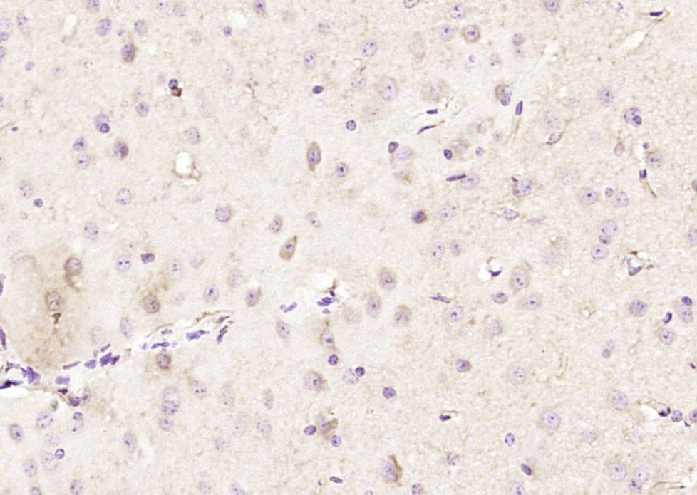
. ADH5 was detected in paraffin-embedded section of human liver cancer tissues. Heat mediated antigen retrieval was performed in citrate buffer (pH6, epitope retrieval solution) for 20 mins. The tissue section was blocked with 10% goat serum. The tissue section was then incubated with 1microg/ml rabbit anti-ADH5 Antibody (RP1111) overnight at 4°C. Biotinylated goat anti-rabbit IgG was used as secondary antibody and incubated for 30 minutes at 37°C. The tissue section was developed using Strepavidin-Biotin-Complex (SABC)(Catalog # SA1022) with DAB as the chromogen.")
. ADH5 was detected in paraffin-embedded section of human mammary cancer tissues. Heat mediated antigen retrieval was performed in citrate buffer (pH6, epitope retrieval solution) for 20 mins. The tissue section was blocked with 10% goat serum. The tissue section was then incubated with 1microg/ml rabbit anti-ADH5 Antibody (RP1111) overnight at 4°C. Biotinylated goat anti-rabbit IgG was used as secondary antibody and incubated for 30 minutes at 37°C. The tissue section was developed using Strepavidin-Biotin-Complex (SABC)(Catalog # SA1022) with DAB as the chromogen.")
. ADH5 was detected in immunocytochemical section of A431 cell. Enzyme antigen retrieval was performed using IHC enzyme antigen retrieval reagent (AR0022) for 15 mins. The cells were blocked with 10% goat serum. And then incubated with 2microg/mL rabbit anti-ADH5 Antibody (RP1111) overnight at 4°C. DyLight®488 Conjugated Goat Anti-Rabbit IgG (BA1127) was used as secondary antibody at 1:100 dilution and incubated for 30 minutes at 37°C. The section was counterstained with DAPI. Visualize using a fluorescence microscope and filter sets appropriate for the label used.")
Figure 1. Western blot analysis of ADH5 using anti-ADH5 antibody (RP1111). Electrophoresis was performed on a 5-20% SDS-PAGE gel at 70V (Stacking gel) / 90V (Resolving gel) for 2-3 hours. The sample well of each lane was loaded with 30 ug of sample under reducing conditions. Lane 1: human HepG2 whole cell lysates, Lane 2: human K562 whole cell lysates, Lane 3: human MOLT4 whole cell lysates, Lane 4: rat liver tissue lysates, Lane 5: mouse liver tissue lysates. After electrophoresis, proteins were transferred to a nitrocellulose membrane at 150 mA for 50-90 minutes. Blocked the membrane with 5% non-fat milk/TBS for 1.5 hour at RT. The membrane was incubated with rabbit anti-ADH5 antigen affinity purified polyclonal antibody (Catalog # RP1111) at 0.5 microg/mL overnight at 4°C, then washed with TBS-0.1%Tween 3 times with 5 minutes each and probed with a goat anti-rabbit IgG-HRP secondary antibody at a dilution of 1:5000 for 1.5 hour at RT. The signal is developed using an Enhanced Chemiluminescent detection (ECL) kit (Catalog # EK1002) with Tanon 5200 system. A specific band was detected for ADH5 at approximately 39 kDa. The expected band size for ADH5 is at 40 kDa.
Anti-Alcohol dehydrogenase class-3 ADH5 Antibody Picoband(r)
RP1111
ApplicationsImmunoFluorescence, Western Blot, ImmunoCytoChemistry, ImmunoHistoChemistry
Product group Antibodies
ReactivityHuman, Mouse, Rat
TargetADH5
Overview
- SupplierBoster Bio
- Product NameAnti-Alcohol dehydrogenase class-3 ADH5 Antibody Picoband(r)
- Delivery Days Customer9
- Application Supplier NoteTested Species: In-house tested species with positive results. By Heat: Boiling the paraffin sections in 10mM citrate buffer, pH6.0, for 20mins is required for the staining of formalin/paraffin sections. Other applications have not been tested. Optimal dilutions should be determined by end users.
- ApplicationsImmunoFluorescence, Western Blot, ImmunoCytoChemistry, ImmunoHistoChemistry
- CertificationResearch Use Only
- ClonalityPolyclonal
- Concentration500 ug/ml
- Gene ID128
- Target nameADH5
- Target descriptionalcohol dehydrogenase 5 (class III), chi polypeptide
- Target synonymsADH-3, ADHX, AMEDS, BMFS7, FALDH, FDH, GSH-FDH, GSNOR, HEL-S-60p, alcohol dehydrogenase class-3, S-(hydroxymethyl)glutathione dehydrogenase, S-nitrosoglutathione reductase, alcohol dehydrogenase (class III), chi polypeptide, alcohol dehydrogenase class chi chain, alcohol dehydrogenase class-III, epididymis secretory sperm binding protein Li 60p, formaldehyde dehydrogenase, glutathione-dependent formaldehyde dehydrogenase
- HostRabbit
- IsotypeIgG
- Protein IDP11766
- Protein NameAlcohol dehydrogenase class-3
- Scientific DescriptionBoster Bio Anti-Alcohol dehydrogenase class-3 ADH5 Antibody catalog # RP1111. Tested in IF, IHC, ICC, WB applications. This antibody reacts with Human, Mouse, Rat. The brand Picoband indicates this is a premium antibody that guarantees superior quality, high affinity, and strong signals with minimal background in Western blot applications. Only our best-performing antibodies are designated as Picoband, ensuring unmatched performance.
- ReactivityHuman, Mouse, Rat
- Storage Instruction-20°C,2°C to 8°C
- UNSPSC12352203
Datasheet
MSDS
Related products
Product group Antibodies
Anti-ADH5 AntibodyA29766
ApplicationsImmunoFluorescence, Western Blot, ImmunoHistoChemistry
ReactivityHuman, Mouse, Rat
- SizePrice
Product group Antibodies
Anti-ADH5 Antibody144-02041
ApplicationsWestern Blot
ReactivityHuman, Mouse
TargetADH5
- SizePrice
Product group Antibodies

ADH5 AntibodyLS-C830603
ApplicationsWestern Blot, ELISA
ReactivityHuman, Mouse, Rat
TargetADH5
- SizePrice
Product group Antibodies
ADH5 Polyclonal AntibodyBS-12448R
ApplicationsFlow Cytometry, ImmunoFluorescence, ELISA, ImmunoCytoChemistry, ImmunoHistoChemistry, ImmunoHistoChemistry Frozen, ImmunoHistoChemistry Paraffin
ReactivityHuman, Mouse, Rat
TargetADH5
- SizePrice
Product group Antibodies
ADH5 AntibodyCSB-PA001357LA01HU
ApplicationsImmunoFluorescence, Western Blot, ELISA, ImmunoHistoChemistry
ReactivityHuman, Rat
TargetADH5
- SizePrice
Product group Antibodies

Goat anti-ADH5EB07612
ApplicationsWestern Blot, ELISA, ImmunoHistoChemistry
ReactivityBovine, Canine, Human, Mouse, Porcine, Rat
TargetADH5
- SizePrice
Product group Antibodies
ADH5 Polyclonal AntibodyCAC14703
ApplicationsImmunoFluorescence, Western Blot, ELISA, ImmunoHistoChemistry
ReactivityRat
TargetADH5
- SizePrice

Product group Antibodies
Anti-ADH5 AntibodyHPA044578
ApplicationsImmunoHistoChemistry
ReactivityHuman
TargetADH5
- SizePrice
Product group Antibodies
ADH5 antibodyGTX55494
ApplicationsWestern Blot
ReactivityHuman
TargetADH5
- SizePrice