


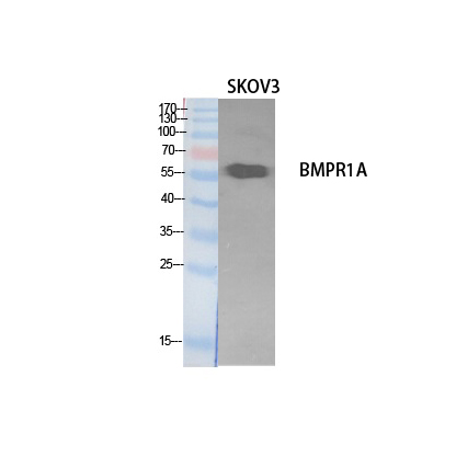
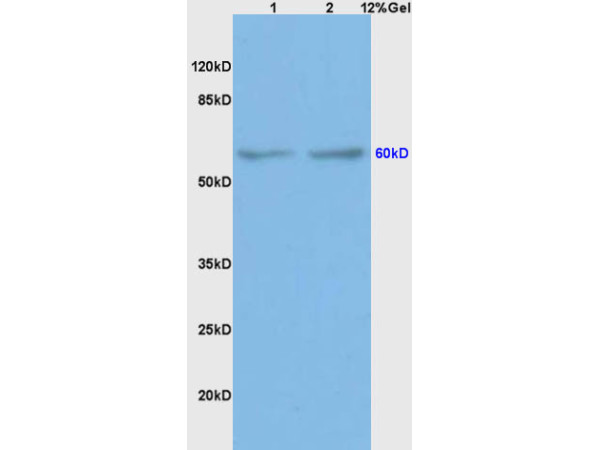
Figure 1. Western blot analysis of BMPR1A using anti-BMPR1A antibody (A01581-1). Electrophoresis was performed on a 5-20% SDS-PAGE gel at 70V (Stacking gel) / 90V (Resolving gel) for 2-3 hours. The sample well of each lane was loaded with 30 ug of sample under reducing conditions. Lane 1: human 293T whole cell lysates, Lane 2: human MCF-7 whole cell lysates, Lane 3: rat skeletal muscle tissue lysates. After electrophoresis, proteins were transferred to a nitrocellulose membrane at 150 mA for 50-90 minutes. Blocked the membrane with 5% non-fat milk/TBS for 1.5 hour at RT. The membrane was incubated with rabbit anti-BMPR1A antigen affinity purified polyclonal antibody (Catalog # A01581-1) at 0.5 microg/mL overnight at 4°C, then washed with TBS-0.1%Tween 3 times with 5 minutes each and probed with a goat anti-rabbit IgG-HRP secondary antibody at a dilution of 1:5000 for 1.5 hour at RT. The signal is developed using an Enhanced Chemiluminescent detection (ECL) kit (Catalog # EK1002) with Tanon 5200 system. A specific band was detected for BMPR1A at approximately 60 kDa. The expected band size for BMPR1A is at 60 kDa.
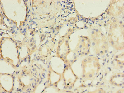
. BMPR1A was detected in a paraffin-embedded section of human breast cancer tissue. Heat mediated antigen retrieval was performed in EDTA buffer (pH 8.0, epitope retrieval solution). The tissue section was blocked with 10% goat serum. The tissue section was then incubated with 2 microg/ml rabbit anti-BMPR1A Antibody (A01581-1) overnight at 4°C. Peroxidase Conjugated Goat Anti-rabbit IgG was used as secondary antibody and incubated for 30 minutes at 37°C. The tissue section was developed using HRP Conjugated Rabbit IgG Super Vision Assay Kit (Catalog # SV0002) with DAB as the chromogen.")
. Overlay histogram showing MCF-7 cells stained with A01581-1 (Blue line). The cells were fixed with 4% paraformaldehyde and blocked with 10% normal goat serum. And then incubated with rabbit anti-BMPR1A Antibody (A01581-1, 1 microg/1x106 cells) for 30 min at 20°C. DyLight®488 conjugated goat anti-rabbit IgG (BA1127, 5-10 microg/1x106 cells) was used as secondary antibody for 30 minutes at 20°C. Isotype control antibody (Green line) was rabbit IgG (1 microg/1x106) used under the same conditions. Unlabelled sample without incubation with primary antibody and secondary antibody (Red line) was used as a blank control.")
Figure 1. Western blot analysis of BMPR1A using anti-BMPR1A antibody (A01581-1). Electrophoresis was performed on a 5-20% SDS-PAGE gel at 70V (Stacking gel) / 90V (Resolving gel) for 2-3 hours. The sample well of each lane was loaded with 30 ug of sample under reducing conditions. Lane 1: human 293T whole cell lysates, Lane 2: human MCF-7 whole cell lysates, Lane 3: rat skeletal muscle tissue lysates. After electrophoresis, proteins were transferred to a nitrocellulose membrane at 150 mA for 50-90 minutes. Blocked the membrane with 5% non-fat milk/TBS for 1.5 hour at RT. The membrane was incubated with rabbit anti-BMPR1A antigen affinity purified polyclonal antibody (Catalog # A01581-1) at 0.5 microg/mL overnight at 4°C, then washed with TBS-0.1%Tween 3 times with 5 minutes each and probed with a goat anti-rabbit IgG-HRP secondary antibody at a dilution of 1:5000 for 1.5 hour at RT. The signal is developed using an Enhanced Chemiluminescent detection (ECL) kit (Catalog # EK1002) with Tanon 5200 system. A specific band was detected for BMPR1A at approximately 60 kDa. The expected band size for BMPR1A is at 60 kDa.
Anti-BMPR1A Antibody Picoband(r)
A01581-1-CARRIER-FREE
ApplicationsFlow Cytometry, Western Blot, ImmunoHistoChemistry
Product group Antibodies
ReactivityHuman, Rat
TargetBMPR1A
Overview
- SupplierBoster Bio
- Product NameAnti-BMPR1A Antibody Picoband(r)
- Delivery Days Customer9
- ApplicationsFlow Cytometry, Western Blot, ImmunoHistoChemistry
- CertificationResearch Use Only
- ClonalityPolyclonal
- Concentration500 ug/ml
- Gene ID657
- Target nameBMPR1A
- Target descriptionbone morphogenetic protein receptor type 1A
- Target synonyms10q23del, ACVRLK3, ALK-3, ALK3, BMPR-1A, CD292, SKR5, bone morphogenetic protein receptor type-1A, BMP type-1A receptor, activin A receptor, type II-like kinase 3, activin receptor-like kinase 3, bone morphogenetic protein receptor, type IA, serine/threonine-protein kinase receptor R5
- HostRabbit
- IsotypeIgG
- Protein IDP36894
- Protein NameBone morphogenetic protein receptor type-1A
- Scientific DescriptionBoster Bio Anti-BMPR1A Antibody Picoband® catalog # A01581-1. Tested in WB, IHC, Flow Cytometry applications. This antibody reacts with Human, Rat. The brand Picoband indicates this is a premium antibody that guarantees superior quality, high affinity, and strong signals with minimal background in Western blot applications. Only our best-performing antibodies are designated as Picoband, ensuring unmatched performance.
- ReactivityHuman, Rat
- Storage Instruction-20°C,2°C to 8°C
- UNSPSC12352203
Related products
Product group Antibodies
ReactivityHuman
TargetBMPR1A
- SizePrice
Product group Antibodies
BMPR1A AntibodyCSB-PA002748LA01HU
ApplicationsImmunoFluorescence, Western Blot, ELISA, ImmunoHistoChemistry
ReactivityHuman
TargetBMPR1A
- SizePrice
Product group Antibodies
Anti-BMPR1A AntibodyA100603
ApplicationsWestern Blot, ELISA
ReactivityHuman
- SizePrice
Product group Antibodies

Anti-BMP receptor type IA [AbD1556]AB00426-2.0
ApplicationsELISA, Neutralisation/Blocking, Other Application
ReactivityHuman
TargetBMPR1A
- SizePrice
Product group Antibodies

Goat anti-BMPR1AEB07197
ApplicationsWestern Blot, ELISA
ReactivityCanine, Human
TargetBMPR1A
- SizePrice
Product group Antibodies

ALK3 / BMPR1A AntibodyLS-C331710
ApplicationsWestern Blot, ImmunoHistoChemistry
ReactivityHuman, Mouse
TargetBMPR1A
- SizePrice
Product group Antibodies
Bmpr1A Polyclonal AntibodyCAC08260
ApplicationsImmunoFluorescence, Western Blot, ELISA, ImmunoHistoChemistry
TargetBMPR1A
- SizePrice
Product group Antibodies
BMPR1A Polyclonal AntibodyBS-1509R
ApplicationsFlow Cytometry, ImmunoFluorescence, Western Blot, ImmunoHistoChemistry, ImmunoHistoChemistry Frozen, ImmunoHistoChemistry Paraffin
ReactivityHuman, Mouse, Rat
TargetBMPR1A
- SizePrice
Product group Antibodies
BMPR1A antibodyGTX113140
ApplicationsWestern Blot
ReactivityHuman
TargetBMPR1A
- SizePrice