


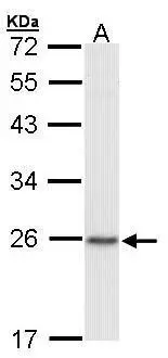
Figure 1. Western blot analysis of CBX1/HP1 beta using anti-CBX1/HP1 beta antibody (A04206-2). Electrophoresis was performed on a 5-20% SDS-PAGE gel at 70V (Stacking gel) / 90V (Resolving gel) for 2-3 hours. The sample well of each lane was loaded with 30 ug of sample under reducing conditions. Lane 1: human Hela whole cell lysates, Lane 2: human A431 whole cell lysates, Lane 3: huamn SW620 whole cell lysates, Lane 4: human CACO-2 whole cell lysates, Lane 5: rat brain tissue lysates, Lane 6: rat testis tissue lysates, Lane 7: mouse brain tissue lysates, Lane 8: mouse testis tissue lysates. After electrophoresis, proteins were transferred to a nitrocellulose membrane at 150 mA for 50-90 minutes. Blocked the membrane with 5% non-fat milk/TBS for 1.5 hour at RT. The membrane was incubated with rabbit anti-CBX1/HP1 beta antigen affinity purified polyclonal antibody (Catalog # A04206-2) at 0.25 microg/mL overnight at 4°C, then washed with TBS-0.1%Tween 3 times with 5 minutes each and probed with a goat anti-rabbit IgG-HRP secondary antibody at a dilution of 1:5000 for 1.5 hour at RT. The signal is developed using an Enhanced Chemiluminescent detection (ECL) kit (Catalog # EK1002) with Tanon 5200 system. A specific band was detected for CBX1/HP1 beta at approximately 21 kDa. The expected band size for CBX1/HP1 beta is at 21 kDa.
. CBX1/HP1 beta was detected in a paraffin-embedded section of human lung cancer tissue. Heat mediated antigen retrieval was performed in EDTA buffer (pH 8.0, epitope retrieval solution). The tissue section was blocked with 10% goat serum. The tissue section was then incubated with 2 microg/ml rabbit anti-CBX1/HP1 beta Antibody (A04206-2) overnight at 4°C. Biotinylated goat anti-rabbit IgG was used as secondary antibody and incubated for 30 minutes at 37°C. The tissue section was developed using Strepavidin-Biotin-Complex (SABC) (Catalog # SA1022) with DAB as the chromogen.")
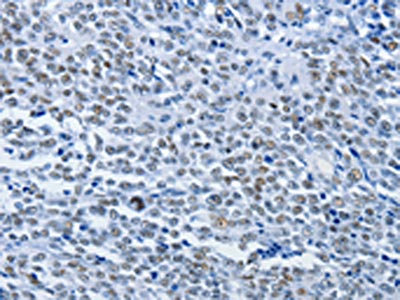
. CBX1/HP1 beta was detected in a paraffin-embedded section of human thyroid papillary carcinoma tissue. Heat mediated antigen retrieval was performed in EDTA buffer (pH 8.0, epitope retrieval solution). The tissue section was blocked with 10% goat serum. The tissue section was then incubated with 2 microg/ml rabbit anti-CBX1/HP1 beta Antibody (A04206-2) overnight at 4°C. Biotinylated goat anti-rabbit IgG was used as secondary antibody and incubated for 30 minutes at 37°C. The tissue section was developed using Strepavidin-Biotin-Complex (SABC) (Catalog # SA1022) with DAB as the chromogen.")
. CBX1/HP1 beta was detected in a paraffin-embedded section of mouse intestines tissue. Heat mediated antigen retrieval was performed in EDTA buffer (pH 8.0, epitope retrieval solution). The tissue section was blocked with 10% goat serum. The tissue section was then incubated with 2 microg/ml rabbit anti-CBX1/HP1 beta Antibody (A04206-2) overnight at 4°C. Biotinylated goat anti-rabbit IgG was used as secondary antibody and incubated for 30 minutes at 37°C. The tissue section was developed using Strepavidin-Biotin-Complex (SABC) (Catalog # SA1022) with DAB as the chromogen.")
. CBX1/HP1 beta was detected in a paraffin-embedded section of rat brain tissue. Heat mediated antigen retrieval was performed in EDTA buffer (pH 8.0, epitope retrieval solution). The tissue section was blocked with 10% goat serum. The tissue section was then incubated with 2 microg/ml rabbit anti-CBX1/HP1 beta Antibody (A04206-2) overnight at 4°C. Biotinylated goat anti-rabbit IgG was used as secondary antibody and incubated for 30 minutes at 37°C. The tissue section was developed using Strepavidin-Biotin-Complex (SABC) (Catalog # SA1022) with DAB as the chromogen.")
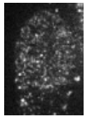
. CBX1/HP1 beta was detected in an immunocytochemical section of CACO-2 cells. Enzyme antigen retrieval was performed using IHC enzyme antigen retrieval reagent (AR0022) for 15 mins. The cells were blocked with 10% goat serum. And then incubated with 5 microg/mL rabbit anti-CBX1/HP1 beta Antibody (A04206-2) overnight at 4°C. DyLight®594 Conjugated Goat Anti-Rabbit IgG (BA1142) was used as secondary antibody at 1:100 dilution and incubated for 30 minutes at 37°C. The section was counterstained with DAPI. Visualize using a fluorescence microscope and filter sets appropriate for the label used.")
Figure 1. Western blot analysis of CBX1/HP1 beta using anti-CBX1/HP1 beta antibody (A04206-2). Electrophoresis was performed on a 5-20% SDS-PAGE gel at 70V (Stacking gel) / 90V (Resolving gel) for 2-3 hours. The sample well of each lane was loaded with 30 ug of sample under reducing conditions. Lane 1: human Hela whole cell lysates, Lane 2: human A431 whole cell lysates, Lane 3: huamn SW620 whole cell lysates, Lane 4: human CACO-2 whole cell lysates, Lane 5: rat brain tissue lysates, Lane 6: rat testis tissue lysates, Lane 7: mouse brain tissue lysates, Lane 8: mouse testis tissue lysates. After electrophoresis, proteins were transferred to a nitrocellulose membrane at 150 mA for 50-90 minutes. Blocked the membrane with 5% non-fat milk/TBS for 1.5 hour at RT. The membrane was incubated with rabbit anti-CBX1/HP1 beta antigen affinity purified polyclonal antibody (Catalog # A04206-2) at 0.25 microg/mL overnight at 4°C, then washed with TBS-0.1%Tween 3 times with 5 minutes each and probed with a goat anti-rabbit IgG-HRP secondary antibody at a dilution of 1:5000 for 1.5 hour at RT. The signal is developed using an Enhanced Chemiluminescent detection (ECL) kit (Catalog # EK1002) with Tanon 5200 system. A specific band was detected for CBX1/HP1 beta at approximately 21 kDa. The expected band size for CBX1/HP1 beta is at 21 kDa.
Anti-CBX1/HP1 beta Antibody Picoband(r)
A04206-2-CARRIER-FREE
ApplicationsImmunoFluorescence, Western Blot, ELISA, ImmunoHistoChemistry
Product group Antibodies
ReactivityHuman, Mouse, Rat
TargetCBX1
Overview
- SupplierBoster Bio
- Product NameAnti-CBX1/HP1 beta Antibody Picoband(r)
- Delivery Days Customer9
- ApplicationsImmunoFluorescence, Western Blot, ELISA, ImmunoHistoChemistry
- CertificationResearch Use Only
- ClonalityPolyclonal
- Concentration500 ug/ml
- Gene ID10951
- Target nameCBX1
- Target descriptionchromobox 1
- Target synonymsCBX, HP1-BETA, HP1Hs-beta, HP1Hsbeta, Hp1beta, M31, MOD1, p25beta, chromobox protein homolog 1, HP1 beta homolog, chromobox homolog 1 (HP1 beta homolog Drosophila ), heterochromatin protein 1 homolog beta, heterochromatin protein 1-beta, heterochromatin protein p25 beta, modifier 1 protein
- HostRabbit
- IsotypeIgG
- Protein IDP83916
- Protein NameChromobox protein homolog 1
- Scientific DescriptionBoster Bio Anti-CBX1/HP1 Antibody Picoband® catalog # A04206-2. Tested in ELISA, IF, IHC, WB applications. This antibody reacts with Human, Mouse, Rat. The brand Picoband indicates this is a premium antibody that guarantees superior quality, high affinity, and strong signals with minimal background in Western blot applications. Only our best-performing antibodies are designated as Picoband, ensuring unmatched performance.
- ReactivityHuman, Mouse, Rat
- Storage Instruction-20°C,2°C to 8°C
- UNSPSC12352203
Related products
Product group Antibodies
ApplicationsImmunoFluorescence, Western Blot, ChIP Chromatin ImmunoPrecipitation
ReactivityHamster, Human
- SizePrice
Product group Antibodies
Anti-CBX1 Antibody144-65503
ApplicationsWestern Blot, ImmunoHistoChemistry
ReactivityHuman
TargetCBX1
- SizePrice
Product group Antibodies

Anti-CBX1 [RAB-C145]Ab01704-1.1
ApplicationsImmunoFluorescence, ImmunoPrecipitation, ChIP Chromatin ImmunoPrecipitation, ELISA
ReactivityHuman
TargetCBX1
- SizePrice
Product group Antibodies

CBX1 / HP1 Beta AntibodyLS-C814104
ApplicationsWestern Blot
ReactivityBovine, Canine, Chicken, Human, Mouse, Porcine, Rat
TargetCBX1
- SizePrice
Product group Antibodies

Goat anti-CBX1 / HP1-BetaEB06958
ApplicationsWestern Blot, ELISA, ImmunoHistoChemistry
ReactivityBovine, Canine, Human, Mouse, Rat
TargetCBX1
- SizePrice
Product group Antibodies
CBX1 AntibodyCSB-PA712321
ApplicationsELISA, ImmunoHistoChemistry
ReactivityHuman, Mouse
TargetCBX1
- SizePrice

Product group Antibodies
Anti-CBX1-25ulHPA071387
ApplicationsWestern Blot, ImmunoCytoChemistry
ReactivityHuman
- SizePrice
Product group Antibodies
CBX1 / HP1 beta antibodyGTX106418
ApplicationsImmunoFluorescence, Western Blot, ImmunoCytoChemistry, ImmunoHistoChemistry, ImmunoHistoChemistry Paraffin
ReactivityHuman, Mouse
TargetCBX1
- SizePrice
Product group Antibodies
Anti-CBX1 AntibodyCAB2247
ApplicationsWestern Blot, ELISA
ReactivityHuman
TargetCBX1
- SizePrice
Product group Antibodies
ApplicationsChIP Chromatin ImmunoPrecipitation
ReactivityHuman, Mouse
TargetCBX1
- SizePrice