

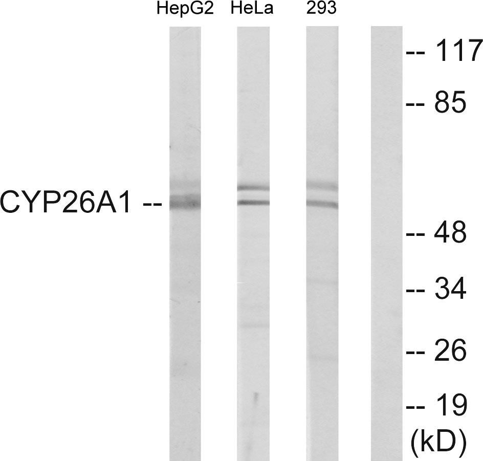


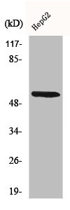
Anti-Cytochrome P450 26A1 Antibody
A94304
ApplicationsWestern Blot, ELISA
Product group Antibodies
ReactivityHuman, Mouse, Rat
Overview
- SupplierAntibodies.com
- Product NameAnti-Cytochrome P450 26A1 Antibody
- Delivery Days Customer7
- ApplicationsWestern Blot, ELISA
- CertificationResearch Use Only
- ClonalityPolyclonal
- ConjugateUnconjugated
- HostRabbit
- IsotypeIgG
- Scientific DescriptionRabbit polyclonal antibody to Cytochrome P450 26A1.
- ReactivityHuman, Mouse, Rat
- UNSPSC12352203
Related products

Product group Antibodies
Anti-CYP26A1 Antibody Picoband(r)A03646-3-CARRIER-FREE
ApplicationsWestern Blot
ReactivityHuman, Mouse, Rat
TargetCYP26A1
- SizePrice
Product group Antibodies

CYP26 / CYP26A1 Antibody (HRP)LS-C682062
ApplicationsELISA
ReactivityZebra Fish
TargetCYP26A1
- SizePrice

Product group Antibodies
CYP26A1 Recombinant AntibodyBSM-54341R
ApplicationsImmunoFluorescence, Western Blot, ImmunoHistoChemistry, ImmunoHistoChemistry Frozen, ImmunoHistoChemistry Paraffin
ReactivityHuman, Mouse, Rat
TargetCYP26A1
- SizePrice
Product group Antibodies
CYP26A1 AntibodyCSB-PA001937
ApplicationsImmunoFluorescence, Western Blot, ELISA
ReactivityHuman, Mouse, Rat
TargetCYP26A1
- SizePrice
Product group Antibodies

Goat anti-CYP26A1EB09041
ApplicationsELISA, ImmunoCytoChemistry, ImmunoHistoChemistry
ReactivityHuman
TargetCYP26A1
- SizePrice
Product group Antibodies
ApplicationsWestern Blot, ImmunoHistoChemistry
ReactivityPorcine
TargetCYP26A1
- SizePrice

Product group Antibodies
CYP26A1 antibodyGTX104961
ApplicationsWestern Blot, ImmunoHistoChemistry, ImmunoHistoChemistry Paraffin
ReactivityHuman, Mouse
TargetCYP26A1
- SizePrice
Product group Antibodies
TargetCYP26A1
- SizePrice
Product group Antibodies
ApplicationsImmunoFluorescence, Western Blot, ELISA, ImmunoCytoChemistry, ImmunoHistoChemistry, ImmunoHistoChemistry Paraffin
ReactivityHuman
TargetCYP26A1
- SizePrice