


CYP26A1 antibody detects CYP26A1 protein at cytoplasm by immunohistochemical analysis. Sample: Paraffin-embedded mouse brain. CYP26A1 stained by CYP26A1 antibody (GTX104961) diluted at 1:500. Antigen Retrieval: Citrate buffer, pH 6.0, 15 min
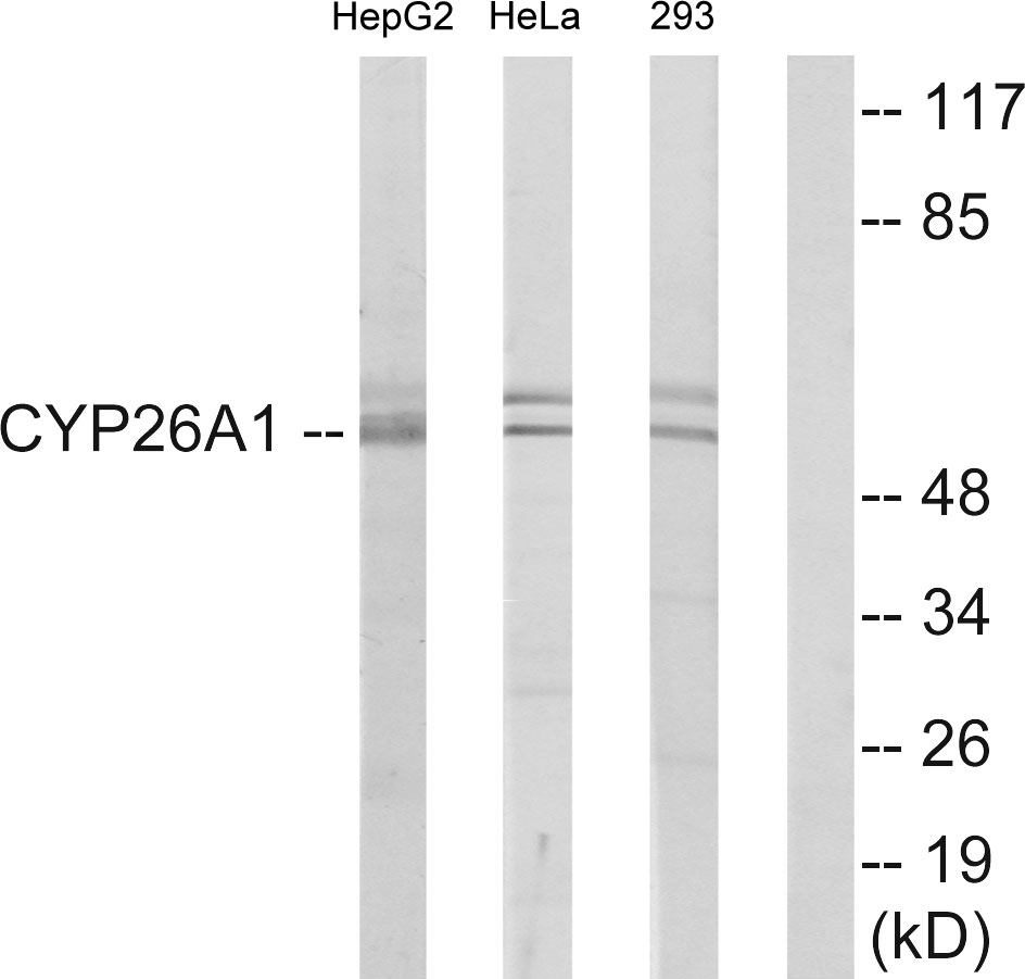
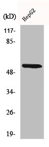
 was separated by 10% SDS-PAGE, and the membrane was blotted with CYP26A1 antibody (GTX104961) diluted at 1:1000. The HRP-conjugated anti-rabbit IgG antibody (GTX213110-01) was used to detect the primary antibody.")
 was separated by 10% SDS-PAGE, and the membrane was blotted with CYP26A1 antibody (GTX104961) diluted at 1:1000. The HRP-conjugated anti-rabbit IgG antibody (GTX213110-01) was used to detect the primary antibody.")
 were separated by 10% SDS-PAGE, and the membrane was blotted with CYP26A1 antibody (GTX104961) diluted at 1:1000. The HRP-conjugated anti-rabbit IgG antibody (GTX213110-01) was used to detect the primary antibody.")
CYP26A1 antibody detects CYP26A1 protein at cytoplasm by immunohistochemical analysis. Sample: Paraffin-embedded mouse brain. CYP26A1 stained by CYP26A1 antibody (GTX104961) diluted at 1:500. Antigen Retrieval: Citrate buffer, pH 6.0, 15 min
CYP26A1 antibody
GTX104961
ApplicationsWestern Blot, ImmunoHistoChemistry, ImmunoHistoChemistry Paraffin
Product group Antibodies
ReactivityHuman, Mouse
TargetCYP26A1
Overview
- SupplierGeneTex
- Product NameCYP26A1 antibody
- Delivery Days Customer9
- Application Supplier NoteWB: 1:500-1:3000. IHC-P: 1:100-1:1000. *Optimal dilutions/concentrations should be determined by the researcher.Not tested in other applications.
- ApplicationsWestern Blot, ImmunoHistoChemistry, ImmunoHistoChemistry Paraffin
- CertificationResearch Use Only
- ClonalityPolyclonal
- Concentration1.33 mg/ml
- ConjugateUnconjugated
- Gene ID1592
- Target nameCYP26A1
- Target descriptioncytochrome P450 family 26 subfamily A member 1
- Target synonymsCP26, CYP26, P450RAI, P450RAI1, cytochrome P450 26A1, P450, retinoic acid-inactivating, 1, cytochrome P450 retinoic acid-inactivating 1, cytochrome P450, family 26, subfamily A, polypeptide 1, cytochrome P450, subfamily XXVIA, polypeptide 1, cytochrome P450RAI, hP450RAI, retinoic acid 4-hydroxylase, retinoic acid-metabolizing cytochrome
- HostRabbit
- IsotypeIgG
- Protein IDO43174
- Protein NameCytochrome P450 26A1
- Scientific DescriptionThis gene encodes a member of the cytochrome P450 superfamily of enzymes. The cytochrome P450 proteins are monooxygenases which catalyze many reactions involved in drug metabolism and synthesis of cholesterol, steroids and other lipids. This endoplasmic reticulum protein acts on retinoids, including all-trans-retinoic acid (RA), with both 4-hydroxylation and 18-hydroxylation activities. This enzyme regulates the cellular level of retinoic acid which is involved in regulation of gene expression in both embryonic and adult tissues. Two alternatively spliced transcript variants of this gene, which encode the distinct isoforms, have been reported. [provided by RefSeq]
- ReactivityHuman, Mouse
- Storage Instruction-20°C or -80°C,2°C to 8°C
- UNSPSC41116161
Datasheet
Related products
Product group Antibodies
ApplicationsWestern Blot, ELISA
ReactivityHuman, Mouse, Rat
- SizePrice

Product group Antibodies
Anti-CYP26A1 Antibody Picoband(r)A03646-3-CARRIER-FREE
ApplicationsWestern Blot
ReactivityHuman, Mouse, Rat
TargetCYP26A1
- SizePrice
Product group Antibodies

CYP26 / CYP26A1 Antibody (HRP)LS-C682062
ApplicationsELISA
ReactivityZebra Fish
TargetCYP26A1
- SizePrice

Product group Antibodies
CYP26A1 Recombinant AntibodyBSM-54341R
ApplicationsImmunoFluorescence, Western Blot, ImmunoHistoChemistry, ImmunoHistoChemistry Frozen, ImmunoHistoChemistry Paraffin
ReactivityHuman, Mouse, Rat
TargetCYP26A1
- SizePrice
Product group Antibodies
CYP26A1 AntibodyCSB-PA001937
ApplicationsImmunoFluorescence, Western Blot, ELISA
ReactivityHuman, Mouse, Rat
TargetCYP26A1
- SizePrice
Product group Antibodies

Goat anti-CYP26A1EB09041
ApplicationsELISA, ImmunoCytoChemistry, ImmunoHistoChemistry
ReactivityHuman
TargetCYP26A1
- SizePrice
Product group Antibodies
ApplicationsWestern Blot, ImmunoHistoChemistry
ReactivityPorcine
TargetCYP26A1
- SizePrice
Product group Antibodies
TargetCYP26A1
- SizePrice
Product group Antibodies
ApplicationsImmunoFluorescence, Western Blot, ELISA, ImmunoCytoChemistry, ImmunoHistoChemistry, ImmunoHistoChemistry Paraffin
ReactivityHuman
TargetCYP26A1
- SizePrice