


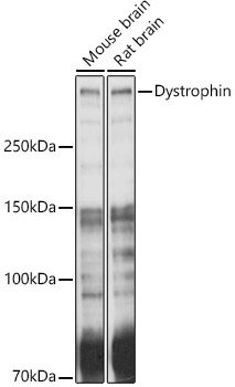
Figure 1. Western blot analysis of Dystrophin using anti-Dystrophin antibody (PB9276). Electrophoresis was performed on a 5-20% SDS-PAGE gel at 70V (Stacking gel) / 90V (Resolving gel) for 2-3 hours. The sample well of each lane was loaded with 50ug of sample under reducing conditions. Lane 1: human HEK293 whole cell lysates, Lane 2: human K562 whole cell lysates, Lane 3: mouse HEPA1-6 whole cell lysates, After Electrophoresis, proteins were transferred to a Nitrocellulose membrane at 150mA for 50-90 minutes. Blocked the membrane with 5% Non-fat Milk/ TBS for 1.5 hour at RT. The membrane was incubated with rabbit anti-Dystrophin antigen affinity purified polyclonal antibody (Catalog # PB9276) at 0.5 microg/mL overnight at 4°C, then washed with TBS-0.1%Tween 3 times with 5 minutes each and probed with a goat anti-rabbit IgG-HRP secondary antibody at a dilution of 1:10000 for 1.5 hour at RT. The signal is developed using an Enhanced Chemiluminescent detection (ECL) kit (Catalog # EK1002) with Tanon 5200 system. A specific band was detected for Dystrophin at approximately 427KD. The expected band size for Dystrophin is at 427KD.
. Dystrophin was detected in paraffin-embedded section of Mouse Brain Tissue. Heat mediated antigen retrieval was performed in citrate buffer (pH6, epitope retrieval solution) for 20 mins. The tissue section was blocked with 10% goat serum. The tissue section was then incubated with 1microg/ml rabbit anti-Dystrophin Antibody (PB9276) overnight at 4°C. Biotinylated goat anti-rabbit IgG was used as secondary antibody and incubated for 30 minutes at 37°C. The tissue section was developed using Strepavidin-Biotin-Complex (SABC)(Catalog # SA1022) with DAB as the chromogen.")
. Dystrophin was detected in paraffin-embedded section of Rat Cardiac Muscle Tissue. Heat mediated antigen retrieval was performed in citrate buffer (pH6, epitope retrieval solution) for 20 mins. The tissue section was blocked with 10% goat serum. The tissue section was then incubated with 1microg/ml rabbit anti-Dystrophin Antibody (PB9276) overnight at 4°C. Biotinylated goat anti-rabbit IgG was used as secondary antibody and incubated for 30 minutes at 37°C. The tissue section was developed using Strepavidin-Biotin-Complex (SABC)(Catalog # SA1022) with DAB as the chromogen.")
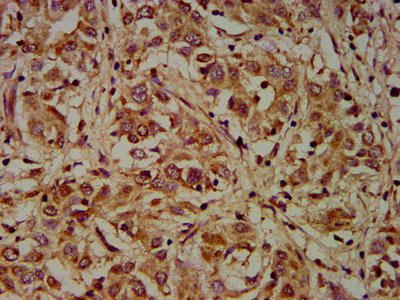
. Dystrophin was detected in paraffin-embedded section of Human Lung Cancer Tissue. Heat mediated antigen retrieval was performed in citrate buffer (pH6, epitope retrieval solution) for 20 mins. The tissue section was blocked with 10% goat serum. The tissue section was then incubated with 1microg/ml rabbit anti-Dystrophin Antibody (PB9276) overnight at 4°C. Biotinylated goat anti-rabbit IgG was used as secondary antibody and incubated for 30 minutes at 37°C. The tissue section was developed using Strepavidin-Biotin-Complex (SABC)(Catalog # SA1022) with DAB as the chromogen.")
. Overlay histogram showing HepG2 cells stained with PB9276 (Blue line).The cells were blocked with 10% normal goat serum. And then incubated with rabbit anti-Dystrophin Antibody (PB9276, 1microg/1x106 cells) for 30 min at 20°C. DyLight?488 conjugated goat anti-rabbit IgG (BA1127, 5-10microg/1x106 cells) was used as secondary antibody for 30 minutes at 20°C. Isotype control antibody (Green line) was rabbit IgG (1microg/1x106) used under the same conditions. Unlabelled sample (Red line) was also used as a control.")
Figure 1. Western blot analysis of Dystrophin using anti-Dystrophin antibody (PB9276). Electrophoresis was performed on a 5-20% SDS-PAGE gel at 70V (Stacking gel) / 90V (Resolving gel) for 2-3 hours. The sample well of each lane was loaded with 50ug of sample under reducing conditions. Lane 1: human HEK293 whole cell lysates, Lane 2: human K562 whole cell lysates, Lane 3: mouse HEPA1-6 whole cell lysates, After Electrophoresis, proteins were transferred to a Nitrocellulose membrane at 150mA for 50-90 minutes. Blocked the membrane with 5% Non-fat Milk/ TBS for 1.5 hour at RT. The membrane was incubated with rabbit anti-Dystrophin antigen affinity purified polyclonal antibody (Catalog # PB9276) at 0.5 microg/mL overnight at 4°C, then washed with TBS-0.1%Tween 3 times with 5 minutes each and probed with a goat anti-rabbit IgG-HRP secondary antibody at a dilution of 1:10000 for 1.5 hour at RT. The signal is developed using an Enhanced Chemiluminescent detection (ECL) kit (Catalog # EK1002) with Tanon 5200 system. A specific band was detected for Dystrophin at approximately 427KD. The expected band size for Dystrophin is at 427KD.
Anti-Dystrophin/DMD Antibody Picoband(r)
PB9276-CARRIER-FREE
ApplicationsFlow Cytometry, Western Blot, ImmunoCytoChemistry, ImmunoHistoChemistry, ImmunoHistoChemistry Frozen
Product group Antibodies
ReactivityBovine, Canine, Chicken, Human, Monkey, Mouse, Rabbit, Rat
TargetDMD
Overview
- SupplierBoster Bio
- Product NameAnti-Dystrophin/DMD Antibody Picoband(r)
- Delivery Days Customer9
- Application Supplier NoteWB: The detection limit for Dystrophin is approximately 0.25ng/lane under reducing conditions. Tested Species: In-house tested species with positive results. By Heat: Boiling the paraffin sections in 10mM citrate buffer, pH6.0, for 20mins is required for the staining of formalin/paraffin sections. Other applications have not been tested. Optimal dilutions should be determined by end users.
- ApplicationsFlow Cytometry, Western Blot, ImmunoCytoChemistry, ImmunoHistoChemistry, ImmunoHistoChemistry Frozen
- CertificationResearch Use Only
- ClonalityPolyclonal
- Concentration500 ug/ml
- Gene ID1756
- Target nameDMD
- Target descriptiondystrophin
- Target synonymsBMD, CMD3B, DXS142, DXS164, DXS206, DXS230, DXS239, DXS268, DXS269, DXS270, DXS272, MRX85, dystrophin, mutant dystrophin
- HostRabbit
- IsotypeIgG
- Protein IDP11532
- Protein NameDystrophin
- Scientific DescriptionBoster Bio Anti-Dystrophin/DMD Antibody Picoband® catalog # PB9276. Tested in Flow Cytometry, IHC, IHC-F, ICC, WB applications. This antibody reacts with Human, Mouse, Rat. The brand Picoband indicates this is a premium antibody that guarantees superior quality, high affinity, and strong signals with minimal background in Western blot applications. Only our best-performing antibodies are designated as Picoband, ensuring unmatched performance.
- ReactivityBovine, Canine, Chicken, Human, Monkey, Mouse, Rabbit, Rat
- Storage Instruction-20°C,2°C to 8°C
- UNSPSC12352203
Related products
Product group Antibodies
DMD AntibodyCSB-PA006963LA01HU
ApplicationsELISA, ImmunoHistoChemistry
ReactivityHuman
TargetDMD
- SizePrice
Product group Antibodies
Anti-DMD Antibody144-63081
ApplicationsImmunoFluorescence, Western Blot, ImmunoHistoChemistry
ReactivityHuman, Mouse, Rat
TargetDMD
- SizePrice
Product group Antibodies

Anti-Dystrophin [133D7-1], Human IgG1, kappaAB04679-10.0
ReactivityHuman
TargetDMD
- SizePrice
Product group Antibodies
Anti-Dystrophin AntibodyA17013
ApplicationsImmunoFluorescence, Western Blot, ImmunoCytoChemistry, ImmunoHistoChemistry
ReactivityHuman, Mouse, Rat
- SizePrice

Product group Antibodies
Anti-DMD AntibodyHPA002725
ApplicationsImmunoHistoChemistry
ReactivityHuman
TargetDMD
- SizePrice
Product group Antibodies

DMD / Dystrophin Antibody (Biotin)LS-C680389
ApplicationsELISA
ReactivityHuman
TargetDMD
- SizePrice
Product group Antibodies
ApplicationsImmunoPrecipitation, Western Blot, ImmunoCytoChemistry, ImmunoHistoChemistry
ReactivityMouse, Porcine, Rat
TargetDMD
- SizePrice
Product group Antibodies
Dystrophin Recombinant AntibodyBSM-61024R
ApplicationsImmunoFluorescence, ImmunoHistoChemistry, ImmunoHistoChemistry Frozen, ImmunoHistoChemistry Paraffin
ReactivityHuman, Mouse, Rat
TargetDMD
- SizePrice
Product group Antibodies
Dystrophin antibody [Dy8/6C5]GTX01868
ApplicationsImmunoHistoChemistry, ImmunoHistoChemistry Frozen
ReactivityCanine, Chicken, Hamster, Human, Mouse, Rabbit, Rat
TargetDMD
- SizePrice