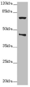
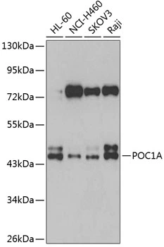
Bio-Connect

Anti-POC1A Antibody

A32090
Antibodies.com
ApplicationsWestern Blot, ImmunoHistoChemistry
Product group Antibodies
Sign in to order and to see your custom pricing.
Large volume orders?
Order with a bulk request

Overview

  • Supplier
    Antibodies.com
  • Product Name
    Anti-POC1A Antibody
  • Delivery Days Customer
    7
  • Applications
    Western Blot, ImmunoHistoChemistry
  • Certification
    Research Use Only
  • Clonality
    Polyclonal
  • Conjugate
    Unconjugated
  • Estimated Purity
    >95%
  • Host
    Rabbit
  • Scientific Description
    Rabbit polyclonal antibody to POC1A
  • UNSPSC
    12352203

Related products

Western blot All lanes: POC1A antibody at 2.76ug/ml + Raji whole cell lysate Secondary Goat polyclonal to rabbit IgG at 1/10000 dilution Predicted band size: 46, 40, 41 kDa Observed band size: 46, 70 kDa
Product group Antibodies
Cusabio
POC1A AntibodyCSB-PA822753ESR1HU
ApplicationsWestern Blot, ELISA, ImmunoHistoChemistry
TargetPOC1A
  • SizePrice
Product group Antibodies
Assay Genie
TargetPOC1A
  • SizePrice
WB analysis of various sample lysates using GTX66024 WDR51A antibody. Dilution : 1:1000 Loading : 25microg per lane
Product group Antibodies
GeneTex
ApplicationsWestern Blot
TargetPOC1A
  • SizePrice
Immunohistochemical staining of human testis shows strong granular cytoplasmic positivity in Leydig cells.
Product group Antibodies
Atlas Antibodies
ApplicationsWestern Blot, ImmunoHistoChemistry
TargetPOC1A
  • SizePrice
Product group Antibodies
RayBiotech
ApplicationsWestern Blot, ImmunoHistoChemistry
TargetPOC1A
  • SizePrice
Figure 1. Western blot analysis of POC1A using anti-POC1A antibody (A08694-1). Electrophoresis was performed on a 5-20% SDS-PAGE gel at 70V (Stacking gel) / 90V (Resolving gel) for 2-3 hours. The sample well of each lane was loaded with 30 ug of sample under reducing conditions. Lane 1: human PC-3 whole cell lysates, Lane 2: human THP-1 whole cell lysates, Lane 3: human U251 whole cell lysates, Lane 4: rat testis tissue lysates, Lane 5: rat thymus tissue lysates, Lane 6: mouse testis tissue lysates, Lane 7: mouse thymus tissue lysates. After electrophoresis, proteins were transferred to a nitrocellulose membrane at 150 mA for 50-90 minutes. Blocked the membrane with 5% non-fat milk/TBS for 1.5 hour at RT. The membrane was incubated with rabbit anti-POC1A antigen affinity purified polyclonal antibody (Catalog # A08694-1) at 0.5 microg/mL overnight at 4°C, then washed with TBS-0.1%Tween 3 times with 5 minutes each and probed with a goat anti-rabbit IgG-HRP secondary antibody at a dilution of 1:5000 for 1.5 hour at RT. The signal is developed using an Enhanced Chemiluminescent detection (ECL) kit (Catalog # EK1002) with Tanon 5200 system. A specific band was detected for POC1A at approximately 40 kDa. The expected band size for POC1A is at 45 kDa.
Product group Antibodies
Boster Bio
Anti-POC1A Antibody Picoband(r)A08694-1-CARRIER-FREE
ApplicationsFlow Cytometry, ImmunoFluorescence, Western Blot, ELISA, ImmunoCytoChemistry, ImmunoHistoChemistry
TargetPOC1A
  • SizePrice
Product group Antibodies
LifeSpan BioSciences
ApplicationsImmunoFluorescence
TargetPOC1A
  • SizePrice