


Immunohistochemical staining of human testis shows strong granular cytoplasmic positivity in Leydig cells.
 and POC1A over-expression lysate (Co-expressed with a C-terminal myc-DDK tag (~3.1 kDa) in mammalian HEK293T cells, LY414586).")
Immunohistochemical staining of human testis shows strong granular cytoplasmic positivity in Leydig cells.
Anti-POC1A Antibody
HPA040600
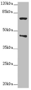
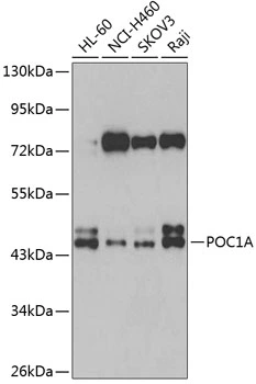
ApplicationsWestern Blot, ImmunoHistoChemistry
Product group Antibodies
ReactivityHuman
TargetPOC1A
Overview
- SupplierAtlas Antibodies
- Product NameAnti-POC1A Antibody
- Delivery Days Customer4
- ApplicationsWestern Blot, ImmunoHistoChemistry
- CertificationResearch Use Only
- ClonalityPolyclonal
- ConjugateUnconjugated
- Gene ID25886
- Target namePOC1A
- Target descriptionPOC1 centriolar protein A
- Target synonymsPIX2, SOFT, WDR51A, POC1 centriolar protein homolog A, WD repeat domain 51A, WD repeat-containing protein 51A, proteome of centriole protein 1A
- HostRabbit
- IsotypeIgG
- Protein IDQ8NBT0
- Protein NamePOC1 centriolar protein homolog A
- Scientific DescriptionRecombinant Protein Epitope Signature Tag (PrEST) antigen sequence
- ReactivityHuman
- Storage Instruction-20°C,2°C to 8°C
- UNSPSC41116161
Datasheet
MSDS
Related products

Product group Antibodies
Anti-POC1A Antibody Picoband(r)A08694-1-CARRIER-FREE
ApplicationsFlow Cytometry, ImmunoFluorescence, Western Blot, ELISA, ImmunoCytoChemistry, ImmunoHistoChemistry
ReactivityHuman, Mouse, Rat
TargetPOC1A
- SizePrice
Product group Antibodies
Anti-POC1A Antibody144-07465
ApplicationsWestern Blot, ImmunoHistoChemistry
ReactivityHuman, Mouse, Rat
TargetPOC1A
- SizePrice
Product group Antibodies
Anti-POC1A AntibodyA32090
ApplicationsWestern Blot, ImmunoHistoChemistry
ReactivityHuman, Mouse, Rat
- SizePrice
Product group Antibodies

POC1A / SOFT AntibodyLS-C748460
ApplicationsImmunoFluorescence
ReactivityHuman
TargetPOC1A
- SizePrice
Product group Antibodies
POC1A AntibodyCSB-PA822753ESR1HU
ApplicationsWestern Blot, ELISA, ImmunoHistoChemistry
ReactivityHuman
TargetPOC1A
- SizePrice
Product group Antibodies
WDR51A antibodyGTX66024
ApplicationsWestern Blot
ReactivityHuman
TargetPOC1A
- SizePrice
Product group Antibodies
Anti-POC1A AntibodyCAB7465
ApplicationsWestern Blot, ELISA
ReactivityHuman
TargetPOC1A
- SizePrice