


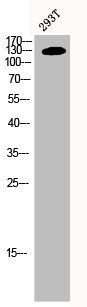
Figure 1. Western blot analysis of PPP1R12A using anti-PPP1R12A antibody (PA1681). Electrophoresis was performed on a 5-20% SDS-PAGE gel at 70V (Stacking gel) / 90V (Resolving gel) for 2-3 hours. The sample well of each lane was loaded with 50ug of sample under reducing conditions. Lane 1: human Hela whole cell lysates, Lane 2: human Jurka7 whole cell lysates, Lane 3: human HEK293 whole cell lysates, Lane 4: monkey COS-7 whole cell lysates, Lane 5: human Raji whole cell lysates, Lane 6: human K562 whole cell lysates, Lane 7: human Caco-2 whole cell lysates, Lane 8: human HepG2 whole cell lysates, After Electrophoresis, proteins were transferred to a Nitrocellulose membrane at 150mA for 50-90 minutes. Blocked the membrane with 5% Non-fat Milk/ TBS for 1.5 hour at RT. The membrane was incubated with rabbit anti-PPP1R12A antigen affinity purified polyclonal antibody (Catalog # PA1681) at 0.5 microg/mL overnight at 4°C, then washed with TBS-0.1%Tween 3 times with 5 minutes each and probed with a goat anti-rabbit IgG-HRP secondary antibody at a dilution of 1:10000 for 1.5 hour at RT. The signal is developed using an Enhanced Chemiluminescent detection (ECL) kit (Catalog # EK1002) with Tanon 5200 system. A specific band was detected for PPP1R12A at approximately 140KD. The expected band size for PPP1R12A is at 115KD.
. Electrophoresis was performed on a 5-20% SDS-PAGE gel at 70V (Stacking gel) / 90V (Resolving gel) for 2-3 hours. The sample well of each lane was loaded with 50ug of sample under reducing conditions. Lane 1: rat brain tissue lysates, Lane 2: rat C6 whole cell lysates, Lane 3: mouse liver tissue lysates, Lane 4: mouse NIH/3T3 whole cell lysates, After Electrophoresis, proteins were transferred to a Nitrocellulose membrane at 150mA for 50-90 minutes. Blocked the membrane with 5% Non-fat Milk/ TBS for 1.5 hour at RT. The membrane was incubated with rabbit anti-PPP1R12A antigen affinity purified polyclonal antibody (Catalog # PA1681) at 0.5 microg/mL overnight at 4°C, then washed with TBS-0.1%Tween 3 times with 5 minutes each and probed with a goat anti-rabbit IgG-HRP secondary antibody at a dilution of 1:10000 for 1.5 hour at RT. The signal is developed using an Enhanced Chemiluminescent detection (ECL) kit (Catalog # EK1002) with Tanon 5200 system. A specific band was detected for PPP1R12A at approximately 140KD. The expected band size for PPP1R12A is at 115KD.")
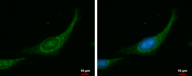
. PPP1R12A was detected in immunocytochemical section of SiHa cells. Enzyme antigen retrieval was performed using IHC enzyme antigen retrieval reagent (AR0022) for 15 mins. The cells were blocked with 10% goat serum. And then incubated with 2microg/mL rabbit anti-PPP1R12A Antibody (PA1681) overnight at 4°C. DyLight?488 Conjugated Goat Anti-Rabbit IgG (BA1127) was used as secondary antibody at 1:100 dilution and incubated for 30 minutes at 37°C. The section was counterstained with DAPI. Visualize using a fluorescence microscope and filter sets appropriate for the label used.")
Figure 1. Western blot analysis of PPP1R12A using anti-PPP1R12A antibody (PA1681). Electrophoresis was performed on a 5-20% SDS-PAGE gel at 70V (Stacking gel) / 90V (Resolving gel) for 2-3 hours. The sample well of each lane was loaded with 50ug of sample under reducing conditions. Lane 1: human Hela whole cell lysates, Lane 2: human Jurka7 whole cell lysates, Lane 3: human HEK293 whole cell lysates, Lane 4: monkey COS-7 whole cell lysates, Lane 5: human Raji whole cell lysates, Lane 6: human K562 whole cell lysates, Lane 7: human Caco-2 whole cell lysates, Lane 8: human HepG2 whole cell lysates, After Electrophoresis, proteins were transferred to a Nitrocellulose membrane at 150mA for 50-90 minutes. Blocked the membrane with 5% Non-fat Milk/ TBS for 1.5 hour at RT. The membrane was incubated with rabbit anti-PPP1R12A antigen affinity purified polyclonal antibody (Catalog # PA1681) at 0.5 microg/mL overnight at 4°C, then washed with TBS-0.1%Tween 3 times with 5 minutes each and probed with a goat anti-rabbit IgG-HRP secondary antibody at a dilution of 1:10000 for 1.5 hour at RT. The signal is developed using an Enhanced Chemiluminescent detection (ECL) kit (Catalog # EK1002) with Tanon 5200 system. A specific band was detected for PPP1R12A at approximately 140KD. The expected band size for PPP1R12A is at 115KD.
Anti-PPP1R12A Antibody
PA1681
ApplicationsImmunoFluorescence, Western Blot, ImmunoCytoChemistry
Product group Antibodies
ReactivityChicken, Human, Monkey, Mouse, Rat
TargetPPP1R12A
Overview
- SupplierBoster Bio
- Product NameAnti-PPP1R12A Antibody
- Delivery Days Customer9
- Application Supplier NoteTested Species: In-house tested species with positive results. Predicted Species: Species predicted to be fit for the product based on sequence similarities. Other applications have not been tested. Optimal dilutions should be determined by end users.
- ApplicationsImmunoFluorescence, Western Blot, ImmunoCytoChemistry
- Applications SupplierWB
- CertificationResearch Use Only
- ClonalityPolyclonal
- Concentration500 ug/ml
- Gene ID4659
- Target namePPP1R12A
- Target descriptionprotein phosphatase 1 regulatory subunit 12A
- Target synonymsGUBS, M130, MBS, MYPT1, protein phosphatase 1 regulatory subunit 12A, myosin binding subunit, myosin phosphatase, target subunit 1, myosin phosphatase-targeting subunit 1, protein phosphatase 1, regulatory (inhibitor) subunit 12A, protein phosphatase myosin-binding subunit
- HostRabbit
- IsotypeIgG
- Protein IDO14974
- Protein NameProtein phosphatase 1 regulatory subunit 12A
- Scientific DescriptionBoster Bio Anti-Myosin Phosphatase/PPP1R12A Antibody catalog # PA1681. Tested in IF, ICC, WB applications. This antibody reacts with Human, Monkey, Mouse, Rat. The brand Picoband indicates this is a premium antibody that guarantees superior quality, high affinity, and strong signals with minimal background in Western blot applications. Only our best-performing antibodies are designated as Picoband, ensuring unmatched performance.
- ReactivityChicken, Human, Monkey, Mouse, Rat
- Reactivity SupplierHuman, Mouse, Rat, Chicken
- Storage Instruction-20°C,2°C to 8°C
- UNSPSC12352203
References
- Dong Q, Luo Y, Yin Y, et al. RhoA/ROCK1 regulates the mitochondrial dysfunction through Drp1 induced by Porphyromonas gingivalis in endothelial cells. J Cell Mol Med. 2023,27(15):2123-2135. doi: 10.1111/jcmm.17796Read this paper
Datasheet
MSDS
Related products
Product group Antibodies
Anti-PPP1R12A Antibody144-00587
ApplicationsImmunoPrecipitation, Western Blot
ReactivityHuman, Mouse
TargetPPP1R12A
- SizePrice
Product group Antibodies
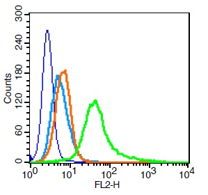
ApplicationsFlow Cytometry, Western Blot
ReactivityBovine, Canine, Chicken, Equine, Human, Mouse, Rabbit, Rat
TargetPPP1R12A
- SizePrice

Product group Antibodies
Anti-Myosin Phosphatase/PPP1R12A Antibody Picoband(r)PB9737-CARRIER-FREE
ApplicationsFlow Cytometry, ImmunoFluorescence, Western Blot, ImmunoCytoChemistry, ImmunoHistoChemistry
ReactivityBovine, Canine, Chicken, Equine, Human, Monkey, Mouse, Rabbit, Rat
TargetPPP1R12A
- SizePrice
Product group Antibodies
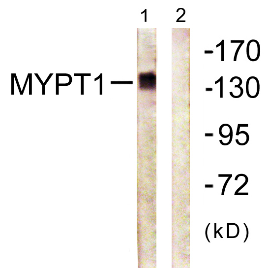
Anti-MYPT1 AntibodyA95830
ApplicationsWestern Blot, ELISA, ImmunoHistoChemistry
ReactivityHuman, Mouse, Rat
- SizePrice
Product group Antibodies
PPP1R12A AntibodyCSB-PA003352
ApplicationsWestern Blot, ELISA, ImmunoHistoChemistry
ReactivityHuman, Monkey, Mouse, Rat
TargetPPP1R12A
- SizePrice

Product group Antibodies
Anti-PPP1R12A AntibodyHPA041296
ApplicationsImmunoCytoChemistry
ReactivityHuman
TargetPPP1R12A
- SizePrice
Product group Antibodies

PPP1R12A / MYPT1 AntibodyLS-C401866
ApplicationsWestern Blot, ELISA
ReactivityHuman, Mouse, Rat
TargetPPP1R12A
- SizePrice
Product group Antibodies
MYPT1 antibodyGTX112344
ApplicationsImmunoFluorescence, ImmunoCytoChemistry
ReactivityHuman
TargetPPP1R12A
- SizePrice