


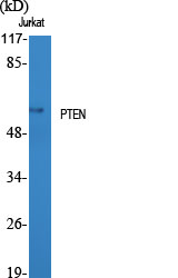
Figure 1. Western blot analysis of PTEN using anti-PTEN antibody (M00006-2). Electrophoresis was performed on a 5-20% SDS-PAGE gel at 70V (Stacking gel) / 90V (Resolving gel) for 2-3 hours. The sample well of each lane was loaded with 30 ug of sample under reducing conditions. Lane 1: human Hela whole cell lysates, Lane 2: human HepG2 whole cell lysates, Lane 3: rat brain tissue lysates, Lane 4: rat PC-12 whole cell lysates, Lane 5: mouse brain tissue lysates, Lane 6: mouse NIH/3T3 whole cell lysates. After electrophoresis, proteins were transferred to a nitrocellulose membrane at 150 mA for 50-90 minutes. Blocked the membrane with 5% non-fat milk/TBS for 1.5 hour at RT. The membrane was incubated with rabbit anti-PTEN antigen affinity purified monoclonal antibody (Catalog # M00006-2) at 1:1000 overnight at 4°C, then washed with TBS-0.1%Tween 3 times with 5 minutes each and probed with a goat anti-rabbit IgG-HRP secondary antibody at a dilution of 1:1000 for 1.5 hour at RT. The signal is developed using an Enhanced Chemiluminescent detection (ECL) kit (Catalog # EK1002) with Tanon 5200 system. A specific band was detected for PTEN at approximately 54 kDa. The expected band size for PTEN is at 47 kDa.


Figure 1. Western blot analysis of PTEN using anti-PTEN antibody (M00006-2). Electrophoresis was performed on a 5-20% SDS-PAGE gel at 70V (Stacking gel) / 90V (Resolving gel) for 2-3 hours. The sample well of each lane was loaded with 30 ug of sample under reducing conditions. Lane 1: human Hela whole cell lysates, Lane 2: human HepG2 whole cell lysates, Lane 3: rat brain tissue lysates, Lane 4: rat PC-12 whole cell lysates, Lane 5: mouse brain tissue lysates, Lane 6: mouse NIH/3T3 whole cell lysates. After electrophoresis, proteins were transferred to a nitrocellulose membrane at 150 mA for 50-90 minutes. Blocked the membrane with 5% non-fat milk/TBS for 1.5 hour at RT. The membrane was incubated with rabbit anti-PTEN antigen affinity purified monoclonal antibody (Catalog # M00006-2) at 1:1000 overnight at 4°C, then washed with TBS-0.1%Tween 3 times with 5 minutes each and probed with a goat anti-rabbit IgG-HRP secondary antibody at a dilution of 1:1000 for 1.5 hour at RT. The signal is developed using an Enhanced Chemiluminescent detection (ECL) kit (Catalog # EK1002) with Tanon 5200 system. A specific band was detected for PTEN at approximately 54 kDa. The expected band size for PTEN is at 47 kDa.
Anti-PTEN Rabbit Monoclonal Antibody
M00006-2
ApplicationsImmunoFluorescence, Western Blot, ImmunoCytoChemistry, ImmunoHistoChemistry
Product group Antibodies
ReactivityHuman, Mouse, Rat
TargetPTEN
Overview
- SupplierBoster Bio
- Product NameAnti-PTEN Rabbit Monoclonal Antibody
- Delivery Days Customer9
- ApplicationsImmunoFluorescence, Western Blot, ImmunoCytoChemistry, ImmunoHistoChemistry
- CertificationResearch Use Only
- ClonalityMonoclonal
- Clone IDHBH-16
- Gene ID5728
- Target namePTEN
- Target descriptionphosphatase and tensin homolog
- Target synonyms10q23del, BZS, CWS1, DEC, GLM2, MHAM, MMAC1, PTEN1, PTENbeta, PTENgama, TEP1, phosphatidylinositol 3,4,5-trisphosphate 3-phosphatase and dual-specificity protein phosphatase PTEN, MMAC1 phosphatase and tensin homolog deleted on chromosome 10, PTEN variant PTEN-K267Rfs*9, PTEN variant PTEN-L247*, PTEN variant PTEN-R234Afs*11, PTENepsilon, inositol polyphosphate 3-phosphatase, mitochondrial PTENalpha, mitochondrial phosphatase and tensin protein alpha, mutated in multiple advanced cancers 1, phosphatase and tensin-like protein, phosphatidylinositol-3,4,5-trisphosphate 3-phosphatase and dual-specificity protein phosphatase PTEN, protein tyrosine phosphatase
- HostRabbit
- IsotypeIgG
- Protein IDP60484
- Protein NamePhosphatidylinositol 3,4,5-trisphosphate 3-phosphatase and dual-specificity protein phosphatase PTEN
- Scientific DescriptionBoster Bio Anti-PTEN Rabbit Monoclonal Antibody catalog # M00006-2. Tested in WB, IHC, ICC/IF applications. This antibody reacts with Human, Mouse, Rat.
- ReactivityHuman, Mouse, Rat
- Storage Instruction-20°C
- UNSPSC12352203
References
- Liu H, Wang X, He K, et al. Oxidized DJ-1 activates the p-IKK/NF-κB/Beclin1 pathway by binding PTEN to induce autophagy and exacerbate myocardial ischemia-reperfusion injury. Eur J Pharmacol. 2024,971:176496. doi: 10.1016/j.ejphar.2024.176496Read this paper
Datasheet
MSDS
Related products
Product group Antibodies
Anti-PTEN AntibodyA96100
ApplicationsWestern Blot, ELISA, ImmunoHistoChemistry
ReactivityHuman, Mouse, Rat
- SizePrice
Product group Antibodies
Anti-PTEN Antibody130-10004
ApplicationsWestern Blot, ELISA
ReactivityHuman
TargetPTEN
- SizePrice

Product group Antibodies
Anti-PTEN AntibodyAMAB91735
ApplicationsWestern Blot, ImmunoHistoChemistry
ReactivityHuman
TargetPTEN
- SizePrice

Product group Antibodies
Anti-PTEN Antibody Picoband(r)A00006-1-CARRIER-FREE
ApplicationsWestern Blot
ReactivityHuman, Mouse, Rat
TargetPTEN
- SizePrice
Product group Antibodies
References
PTEN Polyclonal AntibodyBS-0686R
ApplicationsFlow Cytometry, ImmunoFluorescence, Western Blot, ELISA, ImmunoCytoChemistry, ImmunoHistoChemistry, ImmunoHistoChemistry Frozen, ImmunoHistoChemistry Paraffin
ReactivityMouse, Rat
TargetPTEN
- SizePrice
Product group Antibodies
PTEN AntibodyCSB-PA003874
ApplicationsWestern Blot, ELISA, ImmunoHistoChemistry
ReactivityHuman, Mouse, Rat
TargetPTEN
- SizePrice
Product group Antibodies
References

Goat anti-PTENEB06544
ApplicationsImmunoPrecipitation, Western Blot, ELISA
ReactivityBovine, Canine, Human, Mouse, Porcine, Rat
TargetPTEN
- SizePrice
Product group Antibodies
ApplicationsImmunoPrecipitation, Western Blot, ImmunoCytoChemistry, ImmunoHistoChemistry
ReactivityBovine, Canine, Chicken, Equine, Guinea Pig, Goat, Porcine, Rat, Sheep
TargetPTEN
- SizePrice
Product group Antibodies
PTEN antibodyGTX101025
ApplicationsImmunoFluorescence, Western Blot, ImmunoCytoChemistry, ImmunoHistoChemistry, ImmunoHistoChemistry Paraffin
ReactivityCanine, Feline, Human, Mouse, Porcine, Rat
TargetPTEN
- SizePrice