


Anti-RIP Antibody
A10146
ApplicationsImmunoFluorescence, ImmunoPrecipitation, Western Blot, ImmunoCytoChemistry, ImmunoHistoChemistry
Product group Antibodies
ReactivityHuman, Mouse, Rat
Overview
- SupplierAntibodies.com
- Product NameAnti-RIP Antibody
- Delivery Days Customer7
- ApplicationsImmunoFluorescence, ImmunoPrecipitation, Western Blot, ImmunoCytoChemistry, ImmunoHistoChemistry
- CertificationResearch Use Only
- ClonalityPolyclonal
- ConjugateUnconjugated
- HostRabbit
- IsotypeIgG
- Scientific DescriptionRabbit polyclonal antibody to RIP.
- ReactivityHuman, Mouse, Rat
- UNSPSC12352203
Related products

Product group Antibodies
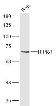
Anti-RIPK1 AntibodyAMAB91705
ApplicationsWestern Blot
ReactivityHuman
TargetRIPK1
- SizePrice
Product group Antibodies

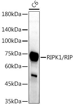
RIPK1 / RIP AntibodyLS-C830300
ApplicationsWestern Blot, ELISA, ImmunoHistoChemistry
ReactivityHuman
TargetRIPK1
- SizePrice
Product group Antibodies
References
RIPK1 Polyclonal AntibodyBS-5805R
ApplicationsFlow Cytometry, ImmunoFluorescence, Western Blot, ELISA, ImmunoCytoChemistry, ImmunoHistoChemistry, ImmunoHistoChemistry Frozen, ImmunoHistoChemistry Paraffin
ReactivityBovine, Equine, Human, Mouse, Porcine, Rabbit, Rat
TargetRIPK1
- SizePrice
Product group Antibodies
RIPK1 AntibodyCSB-PA618785LA01HU
ApplicationsImmunoFluorescence, Western Blot, ELISA
ReactivityHuman, Mouse, Rat
TargetRIPK1
- SizePrice
Product group Antibodies
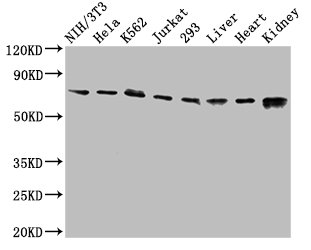
Ripk1 Polyclonal AntibodyCAC09275
ApplicationsImmunoFluorescence, Western Blot, ELISA
ReactivityMouse, Rat
TargetRIPK1
- SizePrice
Product group Antibodies
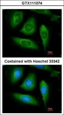
RIP antibodyGTX111074
ApplicationsImmunoFluorescence, Western Blot, ImmunoCytoChemistry
ReactivityHuman
TargetRIPK1
- SizePrice
Product group Antibodies
Anti-RIPK1/RIP AntibodyCAB7414
ApplicationsImmunoFluorescence, ImmunoPrecipitation, Western Blot, ELISA, ImmunoCytoChemistry, ImmunoHistoChemistry, ImmunoHistoChemistry Paraffin
ReactivityHuman
TargetRIPK1
- SizePrice

Product group Antibodies
Anti-RIP/RIPK1 Antibody Picoband(r)PB9116-CARRIER-FREE
ApplicationsWestern Blot
ReactivityHuman
TargetRIPK1
- SizePrice