

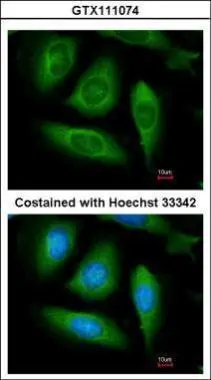
Immunofluorescence analysis of methanol-fixed HeLa, using RIP(GTX111074) antibody at 1:100 dilution.
 A: Molt-4 (GTX27912) 7.5% SDS PAGE GTX111074 diluted at 1:5000")
Immunofluorescence analysis of methanol-fixed HeLa, using RIP(GTX111074) antibody at 1:100 dilution.
RIP antibody
GTX111074
ApplicationsImmunoFluorescence, Western Blot, ImmunoCytoChemistry
Product group Antibodies
ReactivityHuman
TargetRIPK1
Overview
- SupplierGeneTex
- Product NameRIP antibody
- Delivery Days Customer9
- Application Supplier NoteWB: 1:1000-1:10000. ICC/IF: 1:100-1:1000. *Optimal dilutions/concentrations should be determined by the researcher.Not tested in other applications.
- ApplicationsImmunoFluorescence, Western Blot, ImmunoCytoChemistry
- CertificationResearch Use Only
- ClonalityPolyclonal
- Concentration1 mg/ml
- ConjugateUnconjugated
- Gene ID8737
- Target nameRIPK1
- Target descriptionreceptor interacting serine/threonine kinase 1
- Target synonymsAIEFL, IMD57, RIP, RIP-1, RIP1, receptor-interacting serine/threonine-protein kinase 1, cell death protein RIP, receptor (TNFRSF)-interacting serine-threonine kinase 1, receptor-interacting protein 1, receptor-interacting protein kinase 1, serine/threonine-protein kinase RIP
- HostRabbit
- IsotypeIgG
- Protein IDQ13546
- Protein NameReceptor-interacting serine/threonine-protein kinase 1
- Scientific DescriptionEssential adapter molecule for the activation of NF-kappa-B. Following different upstream signals (binding of inflammatory cytokines, stimulation of pathogen recognition receptors, or DNA damage), particular RIPK1-containing complexes are formed, initiating a limited number of cellular responses. Upon TNFA stimulation RIPK1 is recruited to a TRADD-TRAF complex initiated by TNFR1 trimerization. There, it is ubiquitinated via Lys-63-link chains, inducing its association with the IKK complex, and its activation through NEMO binding of polyubiquitin chains.
- ReactivityHuman
- Storage Instruction-20°C or -80°C,2°C to 8°C
- UNSPSC41116161
Datasheet
Related products
Product group Antibodies
Anti-RIP AntibodyA10146
ApplicationsImmunoFluorescence, ImmunoPrecipitation, Western Blot, ImmunoCytoChemistry, ImmunoHistoChemistry
ReactivityHuman, Mouse, Rat
- SizePrice

Product group Antibodies
Anti-RIPK1 AntibodyAMAB91705
ApplicationsWestern Blot
ReactivityHuman
TargetRIPK1
- SizePrice
Product group Antibodies

RIPK1 / RIP AntibodyLS-C830300
ApplicationsWestern Blot, ELISA, ImmunoHistoChemistry
ReactivityHuman
TargetRIPK1
- SizePrice
Product group Antibodies
References
RIPK1 Polyclonal AntibodyBS-5805R
ApplicationsFlow Cytometry, ImmunoFluorescence, Western Blot, ELISA, ImmunoCytoChemistry, ImmunoHistoChemistry, ImmunoHistoChemistry Frozen, ImmunoHistoChemistry Paraffin
ReactivityBovine, Equine, Human, Mouse, Porcine, Rabbit, Rat
TargetRIPK1
- SizePrice
Product group Antibodies
RIPK1 AntibodyCSB-PA618785LA01HU
ApplicationsImmunoFluorescence, Western Blot, ELISA
ReactivityHuman, Mouse, Rat
TargetRIPK1
- SizePrice
Product group Antibodies
Ripk1 Polyclonal AntibodyCAC09275
ApplicationsImmunoFluorescence, Western Blot, ELISA
ReactivityMouse, Rat
TargetRIPK1
- SizePrice
Product group Antibodies
RIP antibodyGTX31389
ApplicationsWestern Blot, ELISA, ImmunoHistoChemistry, ImmunoHistoChemistry Paraffin
ReactivityHuman, Mouse, Rat
TargetRIPK1
- SizePrice
Product group Antibodies
RIP antibodyGTX31684
ApplicationsELISA, ImmunoHistoChemistry, ImmunoHistoChemistry Paraffin
ReactivityHuman
TargetRIPK1
- SizePrice
Product group Antibodies
RIP (phospho Tyr384) antibodyGTX03682
ApplicationsWestern Blot, ELISA, ImmunoHistoChemistry, ImmunoHistoChemistry Paraffin
ReactivityHuman, Mouse
TargetRIPK1
- SizePrice
![Various whole cell extracts (30 μg) were separated by 7.5% SDS-PAGE, and the membrane was blotted with RIP antibody [GT1168] (GTX09128) diluted at 1:500. The HRP-conjugated anti-rabbit IgG antibody (GTX213110-01) was used to detect the primary antibody.](https://www.genetex.com/upload/website/prouct_img/normal/GTX09128/GTX09128_40000000059_20200306_WB_w_23053123_930.webp)
Product group Antibodies
RIP antibody [GT1168]GTX09128
ApplicationsImmunoPrecipitation, Western Blot
ReactivityHuman
TargetRIPK1
- SizePrice