

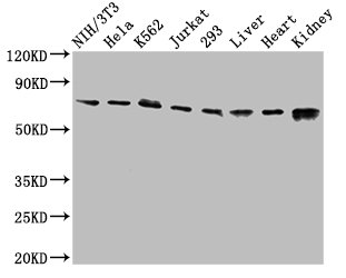
Western Blot Positive WB detected in: NIH/3T3 whole cell lysate, Hela whole cell lysate, K562 whole cell lysate, Jurkat whole cell lysate, 293 whole cell lysate, Rat liver tissue, Mouse heart tissue, Mouse kidney tissue All lanes: RIPK1 antibody at 3.2microg/ml Secondary Goat polyclonal to rabbit IgG at 1/50000 dilution Predicted band size: 76, 71 kDa Observed band size: 76 kDa
.")
Western Blot Positive WB detected in: NIH/3T3 whole cell lysate, Hela whole cell lysate, K562 whole cell lysate, Jurkat whole cell lysate, 293 whole cell lysate, Rat liver tissue, Mouse heart tissue, Mouse kidney tissue All lanes: RIPK1 antibody at 3.2microg/ml Secondary Goat polyclonal to rabbit IgG at 1/50000 dilution Predicted band size: 76, 71 kDa Observed band size: 76 kDa
RIPK1 Antibody
CSB-PA618785LA01HU
ApplicationsImmunoFluorescence, Western Blot, ELISA
Product group Antibodies
ReactivityHuman, Mouse, Rat
TargetRIPK1
Overview
- SupplierCusabio
- Product NameRIPK1 Antibody
- Delivery Days Customer20
- ApplicationsImmunoFluorescence, Western Blot, ELISA
- CertificationResearch Use Only
- ClonalityPolyclonal
- ConjugateUnconjugated
- Gene ID8737
- Target nameRIPK1
- Target descriptionreceptor interacting serine/threonine kinase 1
- Target synonymsAIEFL, IMD57, RIP, RIP-1, RIP1, receptor-interacting serine/threonine-protein kinase 1, cell death protein RIP, receptor (TNFRSF)-interacting serine-threonine kinase 1, receptor-interacting protein 1, receptor-interacting protein kinase 1, serine/threonine-protein kinase RIP
- HostRabbit
- IsotypeIgG
- Protein IDQ13546
- Protein NameReceptor-interacting serine/threonine-protein kinase 1
- Scientific DescriptionSerine-threonine kinase which transduces inflammatory and cell-death signals (programmed necrosis) following death receptors ligation, activation of pathogen recognition receptors (PRRs), and DNA damage. Upon activation of TNFR1 by the TNF-alpha family cytokines, TRADD and TRAF2 are recruited to the receptor. Phosphorylates DAB2IP at Ser-728 in a TNF-alpha-dependent manner, and thereby activates the MAP3K5-JNK apoptotic cascade. Ubiquitination by TRAF2 via Lys-63-link chains acts as a critical enhancer of communication with downstream signal transducers in the mitogen-activated protein kinase pathway and the NF-kappa-B pathway, which in turn mediate downstream events including the activation of genes encoding inflammatory molecules. Polyubiquitinated protein binds to IKBKG/NEMO, the regulatory subunit of the IKK complex, a critical event for NF-kappa-B activation. Interaction with other cellular RHIM-containing adapters initiates gene activation and cell death. RIPK1 and RIPK3 association, in particular, forms a necrosis-inducing complex.
- ReactivityHuman, Mouse, Rat
- Storage Instruction-20°C or -80°C
- UNSPSC41116161
Related products
Product group Antibodies
Anti-RIP AntibodyA10146
ApplicationsImmunoFluorescence, ImmunoPrecipitation, Western Blot, ImmunoCytoChemistry, ImmunoHistoChemistry
ReactivityHuman, Mouse, Rat
- SizePrice

Product group Antibodies
Anti-RIPK1 AntibodyAMAB91705
ApplicationsWestern Blot
ReactivityHuman
TargetRIPK1
- SizePrice
Product group Antibodies

RIPK1 / RIP AntibodyLS-C830300
ApplicationsWestern Blot, ELISA, ImmunoHistoChemistry
ReactivityHuman
TargetRIPK1
- SizePrice
Product group Antibodies
References
RIPK1 Polyclonal AntibodyBS-5805R
ApplicationsFlow Cytometry, ImmunoFluorescence, Western Blot, ELISA, ImmunoCytoChemistry, ImmunoHistoChemistry, ImmunoHistoChemistry Frozen, ImmunoHistoChemistry Paraffin
ReactivityBovine, Equine, Human, Mouse, Porcine, Rabbit, Rat
TargetRIPK1
- SizePrice
Product group Antibodies
Ripk1 Polyclonal AntibodyCAC09275
ApplicationsImmunoFluorescence, Western Blot, ELISA
ReactivityMouse, Rat
TargetRIPK1
- SizePrice
Product group Antibodies
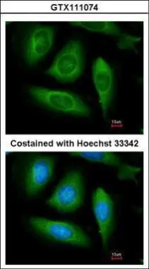
RIP antibodyGTX111074
ApplicationsImmunoFluorescence, Western Blot, ImmunoCytoChemistry
ReactivityHuman
TargetRIPK1
- SizePrice
Product group Antibodies
Anti-RIPK1/RIP AntibodyCAB7414
ApplicationsImmunoFluorescence, ImmunoPrecipitation, Western Blot, ELISA, ImmunoCytoChemistry, ImmunoHistoChemistry, ImmunoHistoChemistry Paraffin
ReactivityHuman
TargetRIPK1
- SizePrice

Product group Antibodies
Anti-RIP/RIPK1 Antibody Picoband(r)PB9116-CARRIER-FREE
ApplicationsWestern Blot
ReactivityHuman
TargetRIPK1
- SizePrice