


Figure 1. Western blot analysis of RIP using anti-RIP antibody (PB9116). Electrophoresis was performed on a 5-20% SDS-PAGE gel at 70V (Stacking gel) / 90V (Resolving gel) for 2-3 hours. Lane 1: recombinant human RIP protein 0.5 ng. After electrophoresis, proteins were transferred to a nitrocellulose membrane at 150 mA for 50-90 minutes. Blocked the membrane with 5% non-fat milk/TBS for 1.5 hour at RT. The membrane was incubated with rabbit anti-RIP antigen affinity purified polyclonal antibody (Catalog # PB9116) at 0.5 microg/mL overnight at 4°C, then washed with TBS-0.1%Tween 3 times with 5 minutes each and probed with a goat anti-rabbit IgG-HRP secondary antibody at a dilution of 1:5000 for 1.5 hour at RT. The signal is developed using an Enhanced Chemiluminescent detection (ECL) kit (Catalog # EK1002) with Tanon 5200 system. A specific band was detected for RIP at approximately 38 kDa. The expected band size for RIP is at 38 kDa.
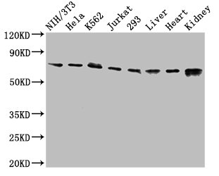
. Electrophoresis was performed on a 5-20% SDS-PAGE gel at 70V (Stacking gel) / 90V (Resolving gel) for 2-3 hours. The sample well of each lane was loaded with 30 ug of sample under reducing conditions. Lane 1: human Jurkat whole cell lysates, Lane 2: human 22RV1 whole cell lysates, Lane 3: human MCF-7 whole cell lysates, Lane 4: human Hela whole cell lysates, Lane 5: human A549 whole cell lysates. After electrophoresis, proteins were transferred to a nitrocellulose membrane at 150 mA for 50-90 minutes. Blocked the membrane with 5% non-fat milk/TBS for 1.5 hour at RT. The membrane was incubated with rabbit anti-RIP antigen affinity purified polyclonal antibody (Catalog # PB9116) at 0.5 microg/mL overnight at 4°C, then washed with TBS-0.1%Tween 3 times with 5 minutes each and probed with a goat anti-rabbit IgG-HRP secondary antibody at a dilution of 1:5000 for 1.5 hour at RT. The signal is developed using an Enhanced Chemiluminescent detection (ECL) kit (Catalog # EK1002) with Tanon 5200 system. A specific band was detected for RIP at approximately 76 kDa. The expected band size for RIP is at 76 kDa.")
Figure 1. Western blot analysis of RIP using anti-RIP antibody (PB9116). Electrophoresis was performed on a 5-20% SDS-PAGE gel at 70V (Stacking gel) / 90V (Resolving gel) for 2-3 hours. Lane 1: recombinant human RIP protein 0.5 ng. After electrophoresis, proteins were transferred to a nitrocellulose membrane at 150 mA for 50-90 minutes. Blocked the membrane with 5% non-fat milk/TBS for 1.5 hour at RT. The membrane was incubated with rabbit anti-RIP antigen affinity purified polyclonal antibody (Catalog # PB9116) at 0.5 microg/mL overnight at 4°C, then washed with TBS-0.1%Tween 3 times with 5 minutes each and probed with a goat anti-rabbit IgG-HRP secondary antibody at a dilution of 1:5000 for 1.5 hour at RT. The signal is developed using an Enhanced Chemiluminescent detection (ECL) kit (Catalog # EK1002) with Tanon 5200 system. A specific band was detected for RIP at approximately 38 kDa. The expected band size for RIP is at 38 kDa.
Anti-RIP/RIPK1 Antibody Picoband(r)
PB9116-CARRIER-FREE
ApplicationsWestern Blot
Product group Antibodies
ReactivityHuman
TargetRIPK1
Overview
- SupplierBoster Bio
- Product NameAnti-RIP/RIPK1 Antibody Picoband(r)
- Delivery Days Customer9
- Application Supplier NoteWB: The detection limit for RIP is approximately 0.25ng/lane under reducing conditions. Tested Species: In-house tested species with positive results. Other applications have not been tested. Optimal dilutions should be determined by end users.
- ApplicationsWestern Blot
- CertificationResearch Use Only
- ClonalityPolyclonal
- Concentration500 ug/ml
- Gene ID8737
- Target nameRIPK1
- Target descriptionreceptor interacting serine/threonine kinase 1
- Target synonymsAIEFL, IMD57, RIP, RIP-1, RIP1, receptor-interacting serine/threonine-protein kinase 1, cell death protein RIP, receptor (TNFRSF)-interacting serine-threonine kinase 1, receptor-interacting protein 1, receptor-interacting protein kinase 1, serine/threonine-protein kinase RIP
- HostRabbit
- IsotypeIgG
- Protein IDQ13546
- Protein NameReceptor-interacting serine/threonine-protein kinase 1
- Scientific DescriptionBoster Bio Anti-RIP/RIPK1 Antibody Picoband® catalog # PB9116. Tested in WB applications. This antibody reacts with Human. The brand Picoband indicates this is a premium antibody that guarantees superior quality, high affinity, and strong signals with minimal background in Western blot applications. Only our best-performing antibodies are designated as Picoband, ensuring unmatched performance.
- ReactivityHuman
- Storage Instruction-20°C,2°C to 8°C
- UNSPSC12352203
Related products
Product group Antibodies
Anti-RIP AntibodyA10146
ApplicationsImmunoFluorescence, ImmunoPrecipitation, Western Blot, ImmunoCytoChemistry, ImmunoHistoChemistry
ReactivityHuman, Mouse, Rat
- SizePrice

Product group Antibodies
Anti-RIPK1 AntibodyAMAB91705
ApplicationsWestern Blot
ReactivityHuman
TargetRIPK1
- SizePrice
Product group Antibodies

RIPK1 / RIP AntibodyLS-C830300
ApplicationsWestern Blot, ELISA, ImmunoHistoChemistry
ReactivityHuman
TargetRIPK1
- SizePrice
Product group Antibodies
References
RIPK1 Polyclonal AntibodyBS-5805R
ApplicationsFlow Cytometry, ImmunoFluorescence, Western Blot, ELISA, ImmunoCytoChemistry, ImmunoHistoChemistry, ImmunoHistoChemistry Frozen, ImmunoHistoChemistry Paraffin
ReactivityBovine, Equine, Human, Mouse, Porcine, Rabbit, Rat
TargetRIPK1
- SizePrice
Product group Antibodies
RIPK1 AntibodyCSB-PA618785LA01HU
ApplicationsImmunoFluorescence, Western Blot, ELISA
ReactivityHuman, Mouse, Rat
TargetRIPK1
- SizePrice
Product group Antibodies
Ripk1 Polyclonal AntibodyCAC09275
ApplicationsImmunoFluorescence, Western Blot, ELISA
ReactivityMouse, Rat
TargetRIPK1
- SizePrice
Product group Antibodies
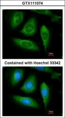
RIP antibodyGTX111074
ApplicationsImmunoFluorescence, Western Blot, ImmunoCytoChemistry
ReactivityHuman
TargetRIPK1
- SizePrice
Product group Antibodies
Anti-RIPK1/RIP AntibodyCAB7414
ApplicationsImmunoFluorescence, ImmunoPrecipitation, Western Blot, ELISA, ImmunoCytoChemistry, ImmunoHistoChemistry, ImmunoHistoChemistry Paraffin
ReactivityHuman
TargetRIPK1
- SizePrice