


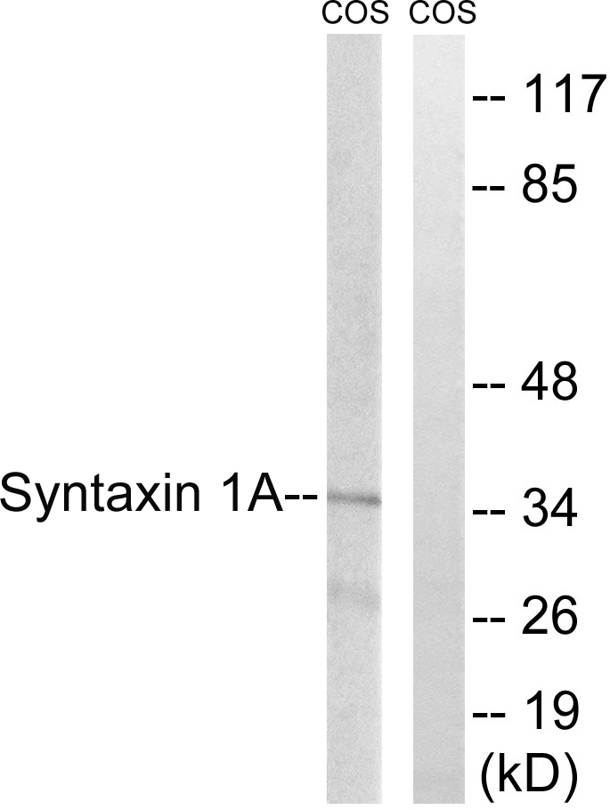
Figure 1. Western blot analysis of Syntaxin 1a using anti-Syntaxin 1a antibody (PB9408). Electrophoresis was performed on a 5-20% SDS-PAGE gel at 70V (Stacking gel) / 90V (Resolving gel) for 2-3 hours. Lane 1: Rat Brain Tissue Lysate at 50ug, Lane 2: Mouse Brain Tissue Lysate at 50ug, Lane 3: U87 Whole Cell Lysate at 40ug. After electrophoresis, proteins were transferred to a nitrocellulose membrane at 150 mA for 50-90 minutes. Blocked the membrane with 5% non-fat milk/TBS for 1.5 hour at RT. The membrane was incubated with rabbit anti-Syntaxin 1a antigen affinity purified polyclonal antibody (Catalog # PB9408) at 0.5 microg/mL overnight at 4°C, then washed with TBS-0.1%Tween 3 times with 5 minutes each and probed with a goat anti-rabbit IgG-HRP secondary antibody at a dilution of 1:5000 for 1.5 hour at RT. The signal is developed using an Enhanced Chemiluminescent detection (ECL) kit (Catalog # EK1002) with Tanon 5200 system. A specific band was detected for Syntaxin 1a at approximately 37 kDa. The expected band size for Syntaxin 1a is at 33 kDa.
Figure 1. Western blot analysis of Syntaxin 1a using anti-Syntaxin 1a antibody (PB9408). Electrophoresis was performed on a 5-20% SDS-PAGE gel at 70V (Stacking gel) / 90V (Resolving gel) for 2-3 hours. Lane 1: Rat Brain Tissue Lysate at 50ug, Lane 2: Mouse Brain Tissue Lysate at 50ug, Lane 3: U87 Whole Cell Lysate at 40ug. After electrophoresis, proteins were transferred to a nitrocellulose membrane at 150 mA for 50-90 minutes. Blocked the membrane with 5% non-fat milk/TBS for 1.5 hour at RT. The membrane was incubated with rabbit anti-Syntaxin 1a antigen affinity purified polyclonal antibody (Catalog # PB9408) at 0.5 microg/mL overnight at 4°C, then washed with TBS-0.1%Tween 3 times with 5 minutes each and probed with a goat anti-rabbit IgG-HRP secondary antibody at a dilution of 1:5000 for 1.5 hour at RT. The signal is developed using an Enhanced Chemiluminescent detection (ECL) kit (Catalog # EK1002) with Tanon 5200 system. A specific band was detected for Syntaxin 1a at approximately 37 kDa. The expected band size for Syntaxin 1a is at 33 kDa.
Anti-Syntaxin 1a/STX1A Antibody Picoband(r)
PB9408-FITC
ApplicationsWestern Blot
Product group Antibodies
ReactivityHamster, Human, Mouse, Rat
TargetSTX1A
Overview
- SupplierBoster Bio
- Product NameAnti-Syntaxin 1a/STX1A Antibody Picoband(r)
- Delivery Days Customer9
- Application Supplier NoteWB: The detection limit for Syntaxin 1a is approximately 0.1ng/lane under reducing conditions. Tested Species: In-house tested species with positive results. Other applications have not been tested. Optimal dilutions should be determined by end users.
- ApplicationsWestern Blot
- CertificationResearch Use Only
- ClonalityPolyclonal
- Concentration500 ug/ml
- ConjugateFITC
- Gene ID6804
- Target nameSTX1A
- Target descriptionsyntaxin 1A
- Target synonymsHPC-1, P35-1, STX1, SYN1A, syntaxin-1A, neuron-specific antigen HPC-1, syntaxin 1A (brain)
- HostRabbit
- IsotypeIgG
- Protein IDQ16623
- Protein NameSyntaxin-1A
- Scientific DescriptionBoster Bio Anti-Syntaxin 1a/STX1A Antibody Picoband® catalog # PB9408. Tested in WB applications. This antibody reacts with Human, Mouse, Rat. The brand Picoband indicates this is a premium antibody that guarantees superior quality, high affinity, and strong signals with minimal background in Western blot applications. Only our best-performing antibodies are designated as Picoband, ensuring unmatched performance.
- ReactivityHamster, Human, Mouse, Rat
- Storage Instruction-20°C,2°C to 8°C
- UNSPSC12352203
Related products
Product group Antibodies
STX1A Polyclonal AntibodyCAC15023
ApplicationsWestern Blot, ELISA
TargetSTX1A
- SizePrice
Product group Antibodies
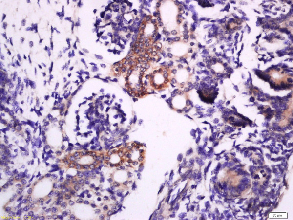
ApplicationsImmunoFluorescence, ELISA, ImmunoCytoChemistry, ImmunoHistoChemistry, ImmunoHistoChemistry Frozen, ImmunoHistoChemistry Paraffin
ReactivityBovine, Canine, Equine, Guinea Pig, Human, Mouse, Porcine, Rat, Sheep
TargetSTX1A
- SizePrice
Product group Antibodies
Anti-STX1A Antibody144-05570
ApplicationsWestern Blot, ImmunoHistoChemistry
ReactivityHuman
TargetSTX1A
- SizePrice
Product group Antibodies
ApplicationsImmunoFluorescence, Western Blot, ELISA, ImmunoHistoChemistry
ReactivityHuman, Mouse, Rat
- SizePrice
Product group Antibodies

ApplicationsWestern Blot, ELISA
ReactivityBovine, Canine, Human, Mouse, Rat
TargetSTX1A
- SizePrice
Product group Antibodies
Syntaxin 1a antibody [N1], N-termGTX106365
ApplicationsImmunoFluorescence, Western Blot, ImmunoCytoChemistry
ReactivityHuman, Rat
TargetSTX1A
- SizePrice
Product group Antibodies
anti-Syntaxin-1A (human), Rabbit Monoclonal (RM367)REV-31-1253-00
ApplicationsWestern Blot, ImmunoHistoChemistry
ReactivityHuman
TargetSTX1A
- SizePrice
Product group Antibodies

STX1A / Syntaxin 1A AntibodyLS-C406049
ApplicationsWestern Blot, ELISA, ImmunoHistoChemistry
ReactivityHuman, Mouse, Rat
TargetSTX1A
- SizePrice