

TNFR-S274 Antibody
ABX026117
ApplicationsFlow Cytometry, Western Blot, ELISA
Product group Antibodies
Overview
- SupplierAbbexa
- Product NameTNFR-S274 Antibody
- Delivery Days Customer12
- ApplicationsFlow Cytometry, Western Blot, ELISA
- CertificationResearch Use Only
- ClonalityPolyclonal
- ConjugateUnconjugated
- HostRabbit
- UNSPSC12352203
Related products
Product group Antibodies
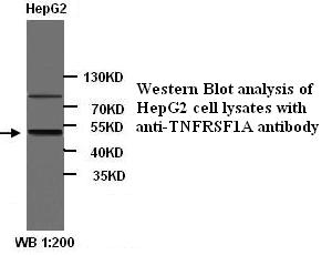
Anti-TNFRSF1A AntibodyA49067
ApplicationsWestern Blot, ELISA
ReactivityHuman
- SizePrice
Product group Antibodies

ApplicationsELISA
ReactivityHuman
TargetTNFRSF1A
- SizePrice

Product group Antibodies
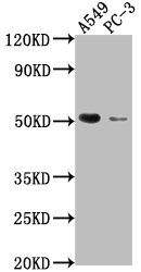
Anti-TNF Receptor I/TNFRSF1A Antibody Picoband(r)A00294-3-CARRIER-FREE
ApplicationsFlow Cytometry, Western Blot, ELISA, ImmunoHistoChemistry
ReactivityHuman, Mouse, Rat
TargetTNFRSF1A
- SizePrice
Product group Antibodies

Goat anti-TNFRSF1A / TNFR1EB11397
ApplicationsWestern Blot, ELISA
ReactivityHuman, Porcine, Rat
TargetTNFRSF1A
- SizePrice
Product group Antibodies
ApplicationsWestern Blot, ImmunoHistoChemistry
ReactivityMouse, Porcine, Rat
TargetTNFRSF1A
- SizePrice
Product group Antibodies
TNFRSF1A AntibodyCSB-PA023977LA01HU
ApplicationsImmunoFluorescence, Western Blot, ELISA, ImmunoHistoChemistry
ReactivityHuman
TargetTNFRSF1A
- SizePrice
Product group Antibodies
References
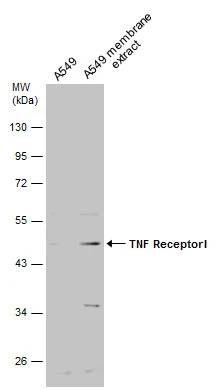
TNFR1 Polyclonal AntibodyBS-2941R
ApplicationsFlow Cytometry, ImmunoFluorescence, Western Blot, ELISA, ImmunoCytoChemistry, ImmunoHistoChemistry, ImmunoHistoChemistry Paraffin
ReactivityBovine, Canine, Equine, Human, Mouse, Porcine, Rabbit, Rat
TargetTNFRSF1A
- SizePrice
Product group Antibodies
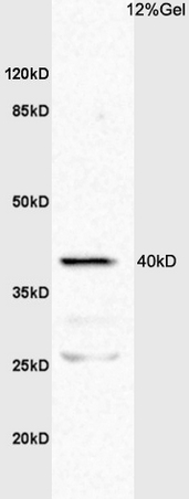
TNF Receptor I antibodyGTX102710
ApplicationsWestern Blot
ReactivityHuman
TargetTNFRSF1A
- SizePrice

Product group Antibodies
Anti-TNFRSF1A AntibodyHPA004102
ApplicationsWestern Blot, ImmunoHistoChemistry
ReactivityHuman
TargetTNFRSF1A
- SizePrice