

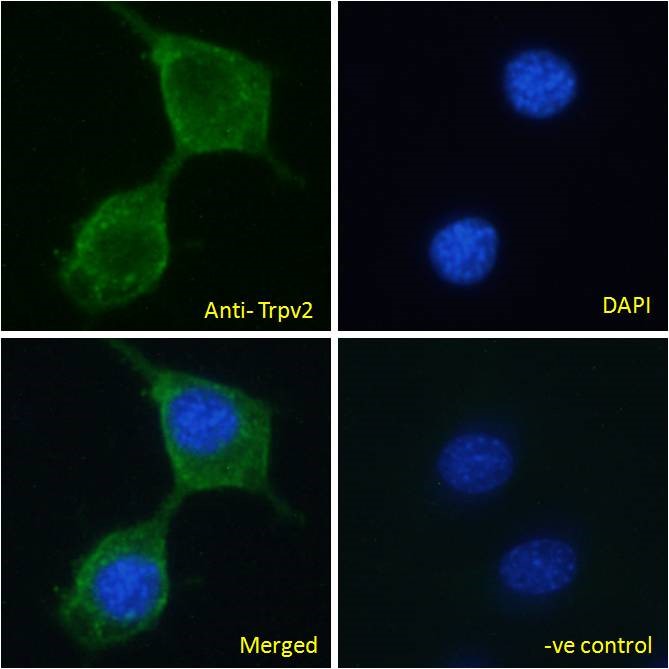
Anti-TRPV2 Antibody
A84565
ApplicationsImmunoFluorescence, ELISA
Product group Antibodies
ReactivityMouse
Overview
- SupplierAntibodies.com
- Product NameAnti-TRPV2 Antibody
- Delivery Days Customer7
- ApplicationsImmunoFluorescence, ELISA
- CertificationResearch Use Only
- ClonalityPolyclonal
- Concentration500 ug/ml
- ConjugateUnconjugated
- HostGoat
- IsotypeIgG
- Scientific DescriptionGoat polyclonal antibody to TRPV2.
- ReactivityMouse
- UNSPSC12352203
Related products

Product group Antibodies
Anti-VRL1/TRPV2 Antibody Picoband(r)A02786-3-CARRIER-FREE
ApplicationsFlow Cytometry, Western Blot, ELISA
ReactivityHuman, Mouse, Rat
TargetTRPV2
- SizePrice
Product group Antibodies

VRL1 / TRPV2 AntibodyLS-C747475
ApplicationsWestern Blot
ReactivityHuman
TargetTRPV2
- SizePrice


Product group Antibodies
Anti-TRPV2 AntibodyHPA044993
ApplicationsImmunoCytoChemistry, ImmunoHistoChemistry
ReactivityHuman
TargetTRPV2
- SizePrice
Product group Antibodies
TRPV2 AntibodyCSB-PA897571LA01HU
ApplicationsImmunoFluorescence, Western Blot, ELISA, ImmunoHistoChemistry
ReactivityHuman, Mouse
TargetTRPV2
- SizePrice
Product group Antibodies
Trpv2 Polyclonal AntibodyCAC11366
ApplicationsImmunoFluorescence, Western Blot, ELISA, ImmunoHistoChemistry
ReactivityMouse
TargetTRPV2
- SizePrice
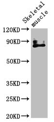
![Unboiled K562 whole cell and membrane extracts (30 μg) were separated by 5% SDS-PAGE, and the membrane was blotted with VRL1 antibody [C2C3], C-term (GTX101868) diluted at 1:500. The HRP-conjugated anti-rabbit IgG antibody (GTX213110-01) was used to detect the primary antibody. (WCE: whole cell extract; ME: membrane extract)](https://www.genetex.com/upload/website/prouct_img/normal/GTX101868/GTX101868_44468_20211126_WB_Fraction_w_23060100_508.webp)
Product group Antibodies
VRL1 antibody [C2C3], C-termGTX101868
ApplicationsWestern Blot, ImmunoHistoChemistry, ImmunoHistoChemistry Paraffin
ReactivityHuman
TargetTRPV2
- SizePrice
Product group Antibodies
Anti-TRPV2 (N-term) Antibody102-21699
ApplicationsWestern Blot
TargetTRPV2
- SizePrice