


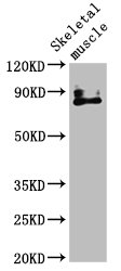
Figure 1. Western blot analysis of VRL1/TRPV2 using anti-VRL1/TRPV2 antibody (A02786-3). Electrophoresis was performed on a 5-20% SDS-PAGE gel at 70V (Stacking gel) / 90V (Resolving gel) for 2-3 hours. The sample well of each lane was loaded with 30ug of sample under reducing conditions. Lane 1: human A549 whole cell lysates, Lane 2: human A431 whole cell lysates, Lane 3: rat brain tissue lysates, Lane 4: mouse brain tissue lysates, Lane 5: mouse heart tissue lysates, Lane 6: mouse lung tissue lysates. After Electrophoresis, proteins were transferred to a Nitrocellulose membrane at 150mA for 50-90 minutes. Blocked the membrane with 5% Non-fat Milk/ TBS for 1.5 hour at RT. The membrane was incubated with rabbit anti-VRL1/TRPV2 antigen affinity purified polyclonal antibody (Catalog # A02786-3) at 0.5 microg/mL overnight at 4°C, then washed with TBS-0.1%Tween 3 times with 5 minutes each and probed with a goat anti-rabbit IgG-HRP secondary antibody at a dilution of 1:5000 for 1.5 hour at RT. The signal is developed using an Enhanced Chemiluminescent detection (ECL) kit (Catalog # EK1002) with Tanon 5200 system. A specific band was detected for VRL1/TRPV2 at approximately 95KD. The expected band size for VRL1/TRPV2 is at 95KD.
. Overlay histogram showing HL-60 cells stained with A02786-3 (Blue line). The cells were fixed with 4% paraformaldehyde and blocked with 10% normal goat serum. And then incubated with rabbit anti-VRL1/TRPV2 Antibody (A02786-3, 1microg/1x106 cells) for 30 min at 20°C. DyLight®488 conjugated goat anti-rabbit IgG (BA1127, 5-10microg/1x106 cells) was used as secondary antibody for 30 minutes at 20°C. Isotype control antibody (Green line) was rabbit IgG (1microg/1x106) used under the same conditions. Unlabelled sample without incubation with primary antibody and secondary antibody (Red line) was used as a blank control.")
Figure 1. Western blot analysis of VRL1/TRPV2 using anti-VRL1/TRPV2 antibody (A02786-3). Electrophoresis was performed on a 5-20% SDS-PAGE gel at 70V (Stacking gel) / 90V (Resolving gel) for 2-3 hours. The sample well of each lane was loaded with 30ug of sample under reducing conditions. Lane 1: human A549 whole cell lysates, Lane 2: human A431 whole cell lysates, Lane 3: rat brain tissue lysates, Lane 4: mouse brain tissue lysates, Lane 5: mouse heart tissue lysates, Lane 6: mouse lung tissue lysates. After Electrophoresis, proteins were transferred to a Nitrocellulose membrane at 150mA for 50-90 minutes. Blocked the membrane with 5% Non-fat Milk/ TBS for 1.5 hour at RT. The membrane was incubated with rabbit anti-VRL1/TRPV2 antigen affinity purified polyclonal antibody (Catalog # A02786-3) at 0.5 microg/mL overnight at 4°C, then washed with TBS-0.1%Tween 3 times with 5 minutes each and probed with a goat anti-rabbit IgG-HRP secondary antibody at a dilution of 1:5000 for 1.5 hour at RT. The signal is developed using an Enhanced Chemiluminescent detection (ECL) kit (Catalog # EK1002) with Tanon 5200 system. A specific band was detected for VRL1/TRPV2 at approximately 95KD. The expected band size for VRL1/TRPV2 is at 95KD.
Anti-VRL1/TRPV2 Antibody Picoband(r)
A02786-3-CARRIER-FREE
ApplicationsFlow Cytometry, Western Blot, ELISA
Product group Antibodies
ReactivityHuman, Mouse, Rat
TargetTRPV2
Overview
- SupplierBoster Bio
- Product NameAnti-VRL1/TRPV2 Antibody Picoband(r)
- Delivery Days Customer9
- ApplicationsFlow Cytometry, Western Blot, ELISA
- CertificationResearch Use Only
- ClonalityPolyclonal
- Concentration500 ug/ml
- Gene ID51393
- Target nameTRPV2
- Target descriptiontransient receptor potential cation channel subfamily V member 2
- Target synonymsVRL, VRL-1, VRL1, transient receptor potential cation channel subfamily V member 2, OTRPC2, osm-9-like TRP channel 2, vanilloid receptor-like protein 1
- HostRabbit
- IsotypeIgG
- Protein IDQ9Y5S1
- Protein NameTransient receptor potential cation channel subfamily V member 2
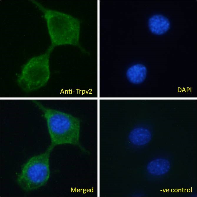
- Scientific DescriptionBoster Bio Anti-VRL1/TRPV2 Antibody Picoband® catalog # A02786-3. Tested in ELISA, Flow Cytometry, WB applications. This antibody reacts with Human, Mouse, Rat. The brand Picoband indicates this is a premium antibody that guarantees superior quality, high affinity, and strong signals with minimal background in Western blot applications. Only our best-performing antibodies are designated as Picoband, ensuring unmatched performance.
- ReactivityHuman, Mouse, Rat
- Storage Instruction-20°C,2°C to 8°C
- UNSPSC12352203
Related products
Product group Antibodies
Anti-TRPV2 AntibodyA84565
ApplicationsImmunoFluorescence, ELISA
ReactivityMouse
- SizePrice
Product group Antibodies

VRL1 / TRPV2 AntibodyLS-C747475
ApplicationsWestern Blot
ReactivityHuman
TargetTRPV2
- SizePrice


Product group Antibodies
Anti-TRPV2 AntibodyHPA044993
ApplicationsImmunoCytoChemistry, ImmunoHistoChemistry
ReactivityHuman
TargetTRPV2
- SizePrice
Product group Antibodies
TRPV2 AntibodyCSB-PA897571LA01HU
ApplicationsImmunoFluorescence, Western Blot, ELISA, ImmunoHistoChemistry
ReactivityHuman, Mouse
TargetTRPV2
- SizePrice
Product group Antibodies
Trpv2 Polyclonal AntibodyCAC11366
ApplicationsImmunoFluorescence, Western Blot, ELISA, ImmunoHistoChemistry
ReactivityMouse
TargetTRPV2
- SizePrice
![Unboiled K562 whole cell and membrane extracts (30 μg) were separated by 5% SDS-PAGE, and the membrane was blotted with VRL1 antibody [C2C3], C-term (GTX101868) diluted at 1:500. The HRP-conjugated anti-rabbit IgG antibody (GTX213110-01) was used to detect the primary antibody. (WCE: whole cell extract; ME: membrane extract)](https://www.genetex.com/upload/website/prouct_img/normal/GTX101868/GTX101868_44468_20211126_WB_Fraction_w_23060100_508.webp)
Product group Antibodies
VRL1 antibody [C2C3], C-termGTX101868
ApplicationsWestern Blot, ImmunoHistoChemistry, ImmunoHistoChemistry Paraffin
ReactivityHuman
TargetTRPV2
- SizePrice
Product group Antibodies
Anti-TRPV2 (N-term) Antibody102-21699
ApplicationsWestern Blot
TargetTRPV2
- SizePrice