


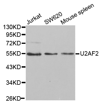
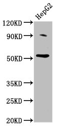
Figure 1. Western blot analysis of U2AF65/U2AF2 using anti-U2AF65/U2AF2 antibody (M03639). Electrophoresis was performed on a 5-20% SDS-PAGE gel at 70V (Stacking gel) / 90V (Resolving gel) for 2-3 hours. The sample well of each lane was loaded with 50ug of sample under reducing conditions. Lane 1: human HEK293 whole cell lysates, Lane 2: human THP-1 whole cell lysates, Lane 3: human U20S whole cell lysates, Lane 4: human Jurkat whole cell lysates, Lane 5: rat PC-12 whole cell lysates, Lane 6: mouse brain tissue lysates, Lane 7: rat brain tissue lysates, Lane 8: mouse RAW264.7 whole cell lysates. After Electrophoresis, proteins were transferred to a Nitrocellulose membrane at 150mA for 50-90 minutes. Blocked the membrane with 5% Non-fat Milk/ TBS for 1.5 hour at RT. The membrane was incubated with mouse anti-U2AF65/U2AF2 antigen affinity purified monoclonal antibody (Catalog # M03639) at 0.25 microg/mL overnight at 4°C, then washed with TBS-0.1%Tween 3 times with 5 minutes each and probed with a goat anti-mouse IgG-HRP secondary antibody at a dilution of 1:10000 for 1.5 hour at RT. The signal is developed using an Enhanced Chemiluminescent detection (ECL) kit (Catalog # EK1001) with Tanon 5200 system. A specific band was detected for U2AF65/U2AF2 at approximately 65KD. The expected band size for U2AF65/U2AF2 is at 65KD.
. Overlay histogram showing A549 cells stained with M03639 (Blue line). To facilitate intracellular staining, cells were fixed with 4% paraformaldehyde and permeabilized with permeabilization buffer. The cells were blocked with 10% normal goat serum. And then incubated with mouse anti-U2AF65/U2AF2 Antibody (M03639, 1microg/1x106 cells) for 30 min at 20°C. DyLight®488 conjugated goat anti-mouse IgG (BA1126, 5-10microg/1x106 cells) was used as secondary antibody for 30 minutes at 20°C. Isotype control antibody (Green line) was mouse IgG (1microg/1x106) used under the same conditions. Unlabelled sample without incubation with primary antibody and secondary antibody (Red line) was used as a blank control.")
. U2AF65/U2AF2 was detected in paraffin-embedded section of human gallbladder adenocarcinoma tissue. Heat mediated antigen retrieval was performed in EDTA buffer (pH8.0, epitope retrieval solution). The tissue section was blocked with 10% goat serum. The tissue section was then incubated with 2microg/ml mouse anti-U2AF65/U2AF2 Antibody (M03639) overnight at 4°C. Biotinylated goat anti-mouse IgG was used as secondary antibody and incubated for 30 minutes at 37°C. The tissue section was developed using Strepavidin-Biotin-Complex (SABC) (Catalog # SA1021) with DAB as the chromogen.")
. U2AF65/U2AF2 was detected in paraffin-embedded section of human adnexal serous adenocarcinoma tissue. Heat mediated antigen retrieval was performed in EDTA buffer (pH8.0, epitope retrieval solution). The tissue section was blocked with 10% goat serum. The tissue section was then incubated with 2microg/ml mouse anti-U2AF65/U2AF2 Antibody (M03639) overnight at 4°C. Biotinylated goat anti-mouse IgG was used as secondary antibody and incubated for 30 minutes at 37°C. The tissue section was developed using Strepavidin-Biotin-Complex (SABC) (Catalog # SA1021) with DAB as the chromogen.")
. U2AF65/U2AF2 was detected in paraffin-embedded section of human colonic adenocarcinoma tissue. Heat mediated antigen retrieval was performed in EDTA buffer (pH8.0, epitope retrieval solution). The tissue section was blocked with 10% goat serum. The tissue section was then incubated with 2microg/ml mouse anti-U2AF65/U2AF2 Antibody (M03639) overnight at 4°C. Biotinylated goat anti-mouse IgG was used as secondary antibody and incubated for 30 minutes at 37°C. The tissue section was developed using Strepavidin-Biotin-Complex (SABC) (Catalog # SA1021) with DAB as the chromogen.")
. U2AF65/U2AF2 was detected in paraffin-embedded section of human colonic adenocarcinoma tissue. Heat mediated antigen retrieval was performed in EDTA buffer (pH8.0, epitope retrieval solution). The tissue section was blocked with 10% goat serum. The tissue section was then incubated with 2microg/ml mouse anti-U2AF65/U2AF2 Antibody (M03639) overnight at 4°C. Biotinylated goat anti-mouse IgG was used as secondary antibody and incubated for 30 minutes at 37°C. The tissue section was developed using Strepavidin-Biotin-Complex (SABC) (Catalog # SA1021) with DAB as the chromogen.")
. U2AF65/U2AF2 was detected in paraffin-embedded section of human lung cancer tissue. Heat mediated antigen retrieval was performed in EDTA buffer (pH8.0, epitope retrieval solution). The tissue section was blocked with 10% goat serum. The tissue section was then incubated with 2microg/ml mouse anti-U2AF65/U2AF2 Antibody (M03639) overnight at 4°C. Biotinylated goat anti-mouse IgG was used as secondary antibody and incubated for 30 minutes at 37°C. The tissue section was developed using Strepavidin-Biotin-Complex (SABC) (Catalog # SA1021) with DAB as the chromogen.")
. U2AF65/U2AF2 was detected in paraffin-embedded section of mouse intestine tissue. Heat mediated antigen retrieval was performed in EDTA buffer (pH8.0, epitope retrieval solution). The tissue section was blocked with 10% goat serum. The tissue section was then incubated with 2microg/ml mouse anti-U2AF65/U2AF2 Antibody (M03639) overnight at 4°C. Biotinylated goat anti-mouse IgG was used as secondary antibody and incubated for 30 minutes at 37°C. The tissue section was developed using Strepavidin-Biotin-Complex (SABC) (Catalog # SA1021) with DAB as the chromogen.")
. U2AF65/U2AF2 was detected in paraffin-embedded section of rat intestine tissue. Heat mediated antigen retrieval was performed in EDTA buffer (pH8.0, epitope retrieval solution). The tissue section was blocked with 10% goat serum. The tissue section was then incubated with 2microg/ml mouse anti-U2AF65/U2AF2 Antibody (M03639) overnight at 4°C. Biotinylated goat anti-mouse IgG was used as secondary antibody and incubated for 30 minutes at 37°C. The tissue section was developed using Strepavidin-Biotin-Complex (SABC) (Catalog # SA1021) with DAB as the chromogen.")
. U2AF65/U2AF2 was detected in immunocytochemical section of A549 cells. Enzyme antigen retrieval was performed using IHC enzyme antigen retrieval reagent (AR0022) for 15 mins. The cells were blocked with 10% goat serum. And then incubated with 5microg/mL mouse anti-U2AF65/U2AF2 Antibody (M03639) overnight at 4°C. DyLight®488 Conjugated Goat Anti-Mouse IgG (BA1126) was used as secondary antibody at 1:100 dilution and incubated for 30 minutes at 37°C. The section was counterstained with DAPI. Visualize using a fluorescence microscope and filter sets appropriate for the label used.")
Figure 1. Western blot analysis of U2AF65/U2AF2 using anti-U2AF65/U2AF2 antibody (M03639). Electrophoresis was performed on a 5-20% SDS-PAGE gel at 70V (Stacking gel) / 90V (Resolving gel) for 2-3 hours. The sample well of each lane was loaded with 50ug of sample under reducing conditions. Lane 1: human HEK293 whole cell lysates, Lane 2: human THP-1 whole cell lysates, Lane 3: human U20S whole cell lysates, Lane 4: human Jurkat whole cell lysates, Lane 5: rat PC-12 whole cell lysates, Lane 6: mouse brain tissue lysates, Lane 7: rat brain tissue lysates, Lane 8: mouse RAW264.7 whole cell lysates. After Electrophoresis, proteins were transferred to a Nitrocellulose membrane at 150mA for 50-90 minutes. Blocked the membrane with 5% Non-fat Milk/ TBS for 1.5 hour at RT. The membrane was incubated with mouse anti-U2AF65/U2AF2 antigen affinity purified monoclonal antibody (Catalog # M03639) at 0.25 microg/mL overnight at 4°C, then washed with TBS-0.1%Tween 3 times with 5 minutes each and probed with a goat anti-mouse IgG-HRP secondary antibody at a dilution of 1:10000 for 1.5 hour at RT. The signal is developed using an Enhanced Chemiluminescent detection (ECL) kit (Catalog # EK1001) with Tanon 5200 system. A specific band was detected for U2AF65/U2AF2 at approximately 65KD. The expected band size for U2AF65/U2AF2 is at 65KD.
Anti-U2AF65/U2AF2 Picoband(r) Antibody (monoclonal, 10F4)
M03639-DYLIGHT488
ApplicationsFlow Cytometry, ImmunoFluorescence, Western Blot, ImmunoCytoChemistry, ImmunoHistoChemistry
Product group Antibodies
ReactivityHuman, Mouse, Rat
TargetU2AF2
Overview
- SupplierBoster Bio
- Product NameAnti-U2AF65/U2AF2 Picoband(r) Antibody (monoclonal, 10F4)
- Delivery Days Customer9
- Application Supplier NoteTested Species: In-house tested species with positive results. Other applications have not been tested. Optimal dilutions should be determined by end users.
- ApplicationsFlow Cytometry, ImmunoFluorescence, Western Blot, ImmunoCytoChemistry, ImmunoHistoChemistry
- CertificationResearch Use Only
- ClonalityMonoclonal
- Clone ID10F4
- Concentration500 ug/ml
- ConjugateDyLight 488
- Gene ID11338
- Target nameU2AF2
- Target descriptionU2 small nuclear RNA auxiliary factor 2
- Target synonymsDEVDFB, U2AF65, splicing factor U2AF 65 kDa subunit, U2 (RNU2) small nuclear RNA auxiliary factor 2, U2 auxiliary factor 65 kDa subunit, U2 small nuclear ribonucleoprotein auxiliary factor (65kD), U2 snRNP auxiliary factor large subunit, hU2AF65
- HostMouse
- IsotypeIgG2b
- Protein IDP26368
- Protein NameSplicing factor U2AF 65 kDa subunit
- Scientific DescriptionBoster Bio Anti-U2AF65/U2AF2 Picoband® Antibody (monoclonal, 10F4) catalog # M03639. Tested in Flow Cytometry, IF, IHC, ICC, WB applications. This antibody reacts with Human, Mouse, Rat. The brand Picoband indicates this is a premium antibody that guarantees superior quality, high affinity, and strong signals with minimal background in Western blot applications. Only our best-performing antibodies are designated as Picoband, ensuring unmatched performance.
- ReactivityHuman, Mouse, Rat
- Storage Instruction-20°C,2°C to 8°C
- UNSPSC12352203
Related products
Product group Antibodies
U2AF2 Polyclonal AntibodyCAC15375
ApplicationsWestern Blot, ELISA
TargetU2AF2
- SizePrice
Product group Antibodies
U2AF2 Recombinant AntibodyBSM-62036R
ApplicationsImmunoFluorescence, Western Blot, ImmunoCytoChemistry, ImmunoHistoChemistry, ImmunoHistoChemistry Frozen, ImmunoHistoChemistry Paraffin
ReactivityHuman, Mouse, Rat
TargetU2AF2
- SizePrice
Product group Antibodies
Anti-U2AF2 AntibodyA30527
ApplicationsImmunoFluorescence, Western Blot, ImmunoHistoChemistry
ReactivityHuman, Mouse, Rat
- SizePrice
Product group Antibodies
Anti-U2AF2 Antibody144-01936
ApplicationsImmunoFluorescence, ImmunoPrecipitation, Western Blot
ReactivityHuman, Mouse
TargetU2AF2
- SizePrice
Product group Antibodies
References
U2AF65 antibody [C1C3]GTX115622
ApplicationsImmunoFluorescence, Western Blot, ImmunoCytoChemistry, ImmunoHistoChemistry, ImmunoHistoChemistry Paraffin
ReactivityHuman, Mouse, Rat
TargetU2AF2
- SizePrice
Product group Antibodies

U2AF2 / U2AF65 AntibodyLS-C331774
ApplicationsImmunoFluorescence, ImmunoPrecipitation, Western Blot, ImmunoHistoChemistry
ReactivityHuman, Mouse
TargetU2AF2
- SizePrice

Product group Antibodies
Anti-U2AF2 AntibodyHPA041943
ApplicationsWestern Blot, ImmunoCytoChemistry
ReactivityHuman
TargetU2AF2
- SizePrice
Product group Antibodies
U2AF2 AntibodyCSB-PA025408LA01HU
ApplicationsWestern Blot, ELISA
ReactivityHuman
TargetU2AF2
- SizePrice