


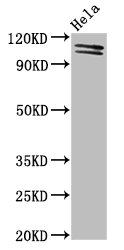
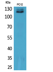
Figure 1. Western blot analysis of UBA1 using anti-UBA1 antibody (PB9904). Electrophoresis was performed on a 5-20% SDS-PAGE gel at 70V (Stacking gel) / 90V (Resolving gel) for 2-3 hours. The sample well of each lane was loaded with 30 ug of sample under reducing conditions. Lane 1: rat liver tissue lysates, Lane 2: mouse liver tissue lysates. Lane 3: mouse testis tissue lysates. Lane 4: HELA whole cell lysates. After electrophoresis, proteins were transferred to a nitrocellulose membrane at 150 mA for 50-90 minutes. Blocked the membrane with 5% non-fat milk/TBS for 1.5 hour at RT. The membrane was incubated with rabbit anti-UBA1 antigen affinity purified polyclonal antibody (Catalog # PB9904) at 0.5 microg/mL overnight at 4°C, then washed with TBS-0.1%Tween 3 times with 5 minutes each and probed with a goat anti-rabbit IgG-HRP secondary antibody at a dilution of 1:5000 for 1.5 hour at RT. The signal is developed using an Enhanced Chemiluminescent detection (ECL) kit (Catalog # EK1002) with Tanon 5200 system. A specific band was detected for UBA1 at approximately 117 kDa. The expected band size for UBA1 is at 118 kDa.
Figure 1. Western blot analysis of UBA1 using anti-UBA1 antibody (PB9904). Electrophoresis was performed on a 5-20% SDS-PAGE gel at 70V (Stacking gel) / 90V (Resolving gel) for 2-3 hours. The sample well of each lane was loaded with 30 ug of sample under reducing conditions. Lane 1: rat liver tissue lysates, Lane 2: mouse liver tissue lysates. Lane 3: mouse testis tissue lysates. Lane 4: HELA whole cell lysates. After electrophoresis, proteins were transferred to a nitrocellulose membrane at 150 mA for 50-90 minutes. Blocked the membrane with 5% non-fat milk/TBS for 1.5 hour at RT. The membrane was incubated with rabbit anti-UBA1 antigen affinity purified polyclonal antibody (Catalog # PB9904) at 0.5 microg/mL overnight at 4°C, then washed with TBS-0.1%Tween 3 times with 5 minutes each and probed with a goat anti-rabbit IgG-HRP secondary antibody at a dilution of 1:5000 for 1.5 hour at RT. The signal is developed using an Enhanced Chemiluminescent detection (ECL) kit (Catalog # EK1002) with Tanon 5200 system. A specific band was detected for UBA1 at approximately 117 kDa. The expected band size for UBA1 is at 118 kDa.
Anti-E1 Ubiquitin Activating Enzyme/UBA1 Antibody Picoband(r)
PB9904
ApplicationsWestern Blot
Product group Antibodies
ReactivityBovine, Equine, Human, Monkey, Mouse, Rabbit, Rat
TargetUBA1
Overview
- SupplierBoster Bio
- Product NameAnti-E1 Ubiquitin Activating Enzyme/UBA1 Antibody Picoband(r)
- Delivery Days Customer9
- Application Supplier NoteTested Species: In-house tested species with positive results. Other applications have not been tested. Optimal dilutions should be determined by end users.
- ApplicationsWestern Blot
- CertificationResearch Use Only
- ClonalityPolyclonal
- Concentration500 ug/ml
- Gene ID7317
- Target nameUBA1
- Target descriptionubiquitin like modifier activating enzyme 1
- Target synonymsA1S9, A1S9T, A1ST, AMCX1, CFAP124, GXP1, POC20, SMAX2, UBA1A, UBE1, UBE1X, VEXAS, ubiquitin-like modifier-activating enzyme 1, A1S9T and BN75 temperature sensitivity complementing, POC20 centriolar protein homolog, UBA1, ubiquitin-activating enzyme E1 homolog A, testicular secretory protein Li 63
- HostRabbit
- IsotypeIgG
- Protein IDP22314
- Protein NameUbiquitin-like modifier-activating enzyme 1
- Scientific DescriptionBoster Bio Anti-E1 Ubiquitin Activating Enzyme/UBA1 Antibody Picoband® catalog # PB9904. Tested in WB applications. This antibody reacts with Human, Mouse, Rat. The brand Picoband indicates this is a premium antibody that guarantees superior quality, high affinity, and strong signals with minimal background in Western blot applications. Only our best-performing antibodies are designated as Picoband, ensuring unmatched performance.
- ReactivityBovine, Equine, Human, Monkey, Mouse, Rabbit, Rat
- Storage Instruction-20°C,2°C to 8°C
- UNSPSC12352203
Datasheet
MSDS
Related products
Product group Antibodies
UBA1 AntibodyCSB-PA00149A0RB
ApplicationsImmunoFluorescence, Western Blot, ELISA
ReactivityHuman
TargetUBA1
- SizePrice
Product group Antibodies
UBA1 Polyclonal AntibodyCAC15035
ApplicationsImmunoFluorescence, Western Blot, ELISA
TargetUBA1
- SizePrice
Product group Antibodies
Anti-UBA1 Antibody144-12359
ApplicationsWestern Blot
ReactivityHuman, Rat
TargetUBA1
- SizePrice
Product group Antibodies
Anti-UBA1 AntibodyA97874
ApplicationsWestern Blot, ELISA
ReactivityHuman, Mouse, Rat
- SizePrice
Product group Antibodies

E1 Ubiquitin AntibodyABX031523
ApplicationsWestern Blot, ELISA, ImmunoHistoChemistry
- SizePrice
Product group Antibodies

UBA1 / UBE1 AntibodyLS-C771550
ApplicationsWestern Blot, ELISA, ImmunoHistoChemistry
ReactivityHuman, Mouse, Rat
TargetUBA1
- SizePrice
Product group Antibodies
TargetUBA1
- SizePrice

Product group Antibodies
ApplicationsImmunoFluorescence, ELISA, ImmunoCytoChemistry, ImmunoHistoChemistry, ImmunoHistoChemistry Frozen, ImmunoHistoChemistry Paraffin
ReactivityHuman, Mouse, Rat
TargetUBA1
- SizePrice