

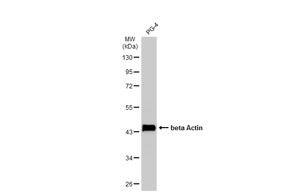
Whole cell extract (30 μg) was separated by 10% SDS-PAGE, and the membrane was blotted with beta Actin antibody (GTX629630) diluted at 1:20000. The HRP-conjugated anti-mouse IgG antibody (GTX213111-01) was used to detect the primary antibody.
![beta Actin antibody [GT5512] detects beta Actin protein at cytoplasm by immunohistochemical analysis. Sample: Paraffin-embedded dog muscle. beta Actin stained by beta Actin antibody [GT5512] (GTX629630) diluted at 1:200. Antigen Retrieval: Citrate buffer, pH 6.0, 15 min](https://www.genetex.com/upload/website/prouct_img/normal/GTX629630/GTX629630_41554_20190201_IHC-P_D_22111423_553.webp "beta Actin antibody [GT5512] detects beta Actin protein at cytoplasm by immunohistochemical analysis. Sample: Paraffin-embedded dog muscle. beta Actin stained by beta Actin antibody [GT5512] (GTX629630) diluted at 1:200. Antigen Retrieval: Citrate buffer, pH 6.0, 15 min")
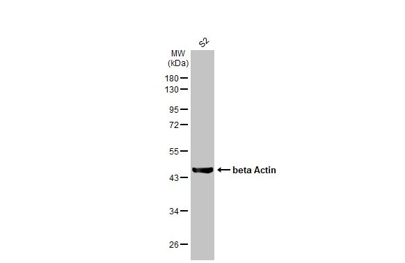
 was separated by 10% SDS-PAGE, and the membrane was blotted with beta Actin antibody (GTX629630) diluted at 1:20000. The HRP-conjugated anti-mouse IgG antibody (GTX213111-01) was used to detect the primary antibody.")
 diluted at 1:500. Blue: Hoechst 33342 staining. Scale bar= 10 μm.")
![beta Actin antibody [GT5512] detects beta Actin protein at cytoskeleton by immunofluorescent analysis. Sample: HeLa cells were fixed in 0.5% Triton X-100 for 1 min, then ice-cold methanol for 5 min. Green: beta Actin protein stained by beta Actin antibody [GT5512] (GTX629630) diluted at 1:500. Blue: Hoechst 33342 staining.](https://www.genetex.com/upload/website/prouct_img/normal/GTX629630/GTX629630_42898_20170719_IFA_w_23061202_239.webp "beta Actin antibody [GT5512] detects beta Actin protein at cytoskeleton by immunofluorescent analysis. Sample: HeLa cells were fixed in 0.5% Triton X-100 for 1 min, then ice-cold methanol for 5 min. Green: beta Actin protein stained by beta Actin antibody [GT5512] (GTX629630) diluted at 1:500. Blue: Hoechst 33342 staining.")
![Beta Actin antibody [GT5512] detects beta Actin protein by western blot analysis. A. 20 μg 293T whole cell lysate/extract B. 10 μg 293T whole cell lysate/extract C. 5 μg 293T whole cell lysate/extract D. 1 μg 293T whole cell lysate/extract 10% SDS-PAGE Beta Actin antibody [GT5512] (GTX629630) dilution: 1:10000 The HRP-conjugated anti-mouse IgG antibody (GTX213111-01) was used to detect the primary antibody.](https://www.genetex.com/upload/website/prouct_img/normal/GTX629630/GTX629630_41379_WB_Sensitivity_w_23061202_154.webp "Beta Actin antibody [GT5512] detects beta Actin protein by western blot analysis. A. 20 μg 293T whole cell lysate/extract B. 10 μg 293T whole cell lysate/extract C. 5 μg 293T whole cell lysate/extract D. 1 μg 293T whole cell lysate/extract 10% SDS-PAGE Beta Actin antibody [GT5512] (GTX629630) dilution: 1:10000 The HRP-conjugated anti-mouse IgG antibody (GTX213111-01) was used to detect the primary antibody.")
![Beta Actin antibody [GT5512] detects beta Actin protein by western blot analysis. A. 30 μg drosophila lysate/extract 10% SDS-PAGE Beta Actin antibody [GT5512] (GTX629630) dilution: 1:1000 The HRP-conjugated anti-mouse IgG antibody (GTX213111-01) was used to detect the primary antibody.](https://www.genetex.com/upload/website/prouct_img/normal/GTX629630/GTX629630_41379_WB_Drosophila_w_23061202_726.webp "Beta Actin antibody [GT5512] detects beta Actin protein by western blot analysis. A. 30 μg drosophila lysate/extract 10% SDS-PAGE Beta Actin antibody [GT5512] (GTX629630) dilution: 1:1000 The HRP-conjugated anti-mouse IgG antibody (GTX213111-01) was used to detect the primary antibody.")
![beta Actin antibody [GT5512] detects beta Actin protein by western blot analysis. A. 30 μg goat blood lysate/extract 10% SDS-PAGE beta Actin antibody [GT5512] (GTX629630) dilution: 1:10000 The HRP-conjugated anti-mouse IgG antibody (GTX213111-01) was used to detect the primary antibody.](https://www.genetex.com/upload/website/prouct_img/normal/GTX629630/GTX629630_41379_WB_Goat_w_23061202_557.webp "beta Actin antibody [GT5512] detects beta Actin protein by western blot analysis. A. 30 μg goat blood lysate/extract 10% SDS-PAGE beta Actin antibody [GT5512] (GTX629630) dilution: 1:10000 The HRP-conjugated anti-mouse IgG antibody (GTX213111-01) was used to detect the primary antibody.")
![Non-transfected (–) and transfected (+) 293T whole cell extracts (10 μg) were separated by 10% SDS-PAGE, and the membrane was blotted with beta Actin antibody [GT5512] (GTX629630) diluted at 1:20000. The HRP-conjugated anti-mouse IgG antibody (GTX213111-01) was used to detect the primary antibody.](https://www.genetex.com/upload/website/prouct_img/normal/GTX629630/GTX629630_41554_20160707_WB_shRNA_watermark_w_23061202_592.webp "Non-transfected (–) and transfected (+) 293T whole cell extracts (10 μg) were separated by 10% SDS-PAGE, and the membrane was blotted with beta Actin antibody [GT5512] (GTX629630) diluted at 1:20000. The HRP-conjugated anti-mouse IgG antibody (GTX213111-01) was used to detect the primary antibody.")
![beta Actin antibody [GT5512] detects beta Actin protein by western blot analysis. A. 30 μg rabbit blood lysate/extract 10% SDS-PAGE beta Actin antibody [GT5512] (GTX629630) dilution: 1:10000 The HRP-conjugated anti-mouse IgG antibody (GTX213111-01) was used to detect the primary antibody.](https://www.genetex.com/upload/website/prouct_img/normal/GTX629630/GTX629630_41379_WB_Rabbit_w_23061202_374.webp "beta Actin antibody [GT5512] detects beta Actin protein by western blot analysis. A. 30 μg rabbit blood lysate/extract 10% SDS-PAGE beta Actin antibody [GT5512] (GTX629630) dilution: 1:10000 The HRP-conjugated anti-mouse IgG antibody (GTX213111-01) was used to detect the primary antibody.")
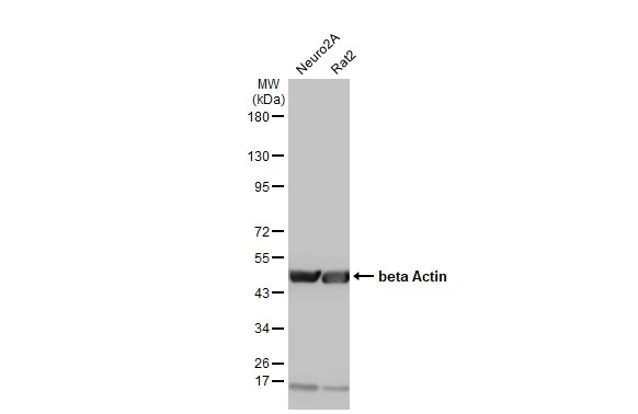
Whole cell extract (30 μg) was separated by 10% SDS-PAGE, and the membrane was blotted with beta Actin antibody (GTX629630) diluted at 1:20000. The HRP-conjugated anti-mouse IgG antibody (GTX213111-01) was used to detect the primary antibody.
beta Actin antibody [GT5512]
GTX629630
ApplicationsImmunoFluorescence, ImmunoPrecipitation, Western Blot, ImmunoCytoChemistry, ImmunoHistoChemistry, ImmunoHistoChemistry Paraffin
Product group Antibodies
ReactivityCanine, Drosophila, Feline, Goat, Human, Monkey, Mouse, Rabbit, Rat, Xenopus, Yeast, Zebra Fish
TargetACTB
Overview
- SupplierGeneTex
- Product Namebeta Actin antibody [GT5512]
- Delivery Days Customer9
- Application Supplier NoteWB: 1:500-1:20000. ICC/IF: 1:100-1:1000. IHC-P: 1:100-1:1000. *Optimal dilutions/concentrations should be determined by the researcher.Not tested in other applications.
- ApplicationsImmunoFluorescence, ImmunoPrecipitation, Western Blot, ImmunoCytoChemistry, ImmunoHistoChemistry, ImmunoHistoChemistry Paraffin
- CertificationResearch Use Only
- ClonalityMonoclonal
- Clone IDGT5512
- Concentration1 mg/ml
- ConjugateUnconjugated
- Gene ID60
- Target nameACTB
- Target descriptionactin beta
- Target synonymsBKRNS, BNS, BRWS1, CSMH, DDS1, PS1TP5BP1, THC8, actin, cytoplasmic 1, I(2)-actin, PS1TP5-binding protein 1, beta cytoskeletal actin
- HostMouse
- IsotypeIgG1
- Protein IDP60709
- Protein NameActin, cytoplasmic 1
- Scientific DescriptionThis gene encodes one of six different actin proteins. Actins are highly conserved proteins that are involved in cell motility, structure, and integrity. This actin is a major constituent of the contractile apparatus and one of the two nonmuscle cytoskeletal actins. [provided by RefSeq]
- ReactivityCanine, Drosophila, Feline, Goat, Human, Monkey, Mouse, Rabbit, Rat, Xenopus, Yeast, Zebra Fish
- Storage Instruction-20°C or -80°C,2°C to 8°C
- UNSPSC12352203
References
- Cermakova K, Tao L, Dejmek M, et al. Reactivation of the G1 enhancer landscape underlies core circuitry addiction to SWI/SNF. Nucleic Acids Res. 2024,52(1):4-21. doi: 10.1093/nar/gkad1081Read this paper
- Sulistyowati E, Huang SE, Cheng TL, et al. Vasculoprotective Potential of Baicalein in Angiotensin II-Infused Abdominal Aortic Aneurysms through Inhibiting Inflammation and Oxidative Stress. Int J Mol Sci. 2023,24(21). doi: 10.3390/ijms242116004Read this paper
- Ballon Romero SS, Fuh LJ, Hung SY, et al. Electroacupuncture exerts prolonged analgesic and neuroprotective effects in a persistent dental pain model induced by multiple dental pulp injuries: GABAergic interneurons-astrocytes interaction. Front Immunol. 2023,14:1213710. doi: 10.3389/fimmu.2023.1213710Read this paper
- Kim HY, Lee J, Kim HJ, et al. PLCγ1 in dopamine neurons critically regulates striatal dopamine release via VMAT2 and synapsin III. Exp Mol Med. 2023,55(11):2357-2375. doi: 10.1038/s12276-023-01104-yRead this paper
- Kadirvelu J, Jacobs SE, Liu R, et al. The E3 ubiquitin ligase RNF216 contains a linear ubiquitin chain-determining-like domain that functions to regulate dendritic arborization and dendritic spine type in hippocampal neurons. bioRxiv. 2023,:pii: 2023.10.19.563080. doi: 10.1101/2023.10.19.563080.Read this paper
- Mercado-Gómez OF, Arriaga-Ávila VS, Vega-García A, et al. Daytime-Restricted Feeding Ameliorates Oxidative Stress by Increasing NRF2 Transcriptional Factor in the Rat Hippocampus in the Pilocarpine-Induced Acute Seizure Model. Brain Sci. 2023,13(10). doi: 10.3390/brainsci13101442Read this paper
- Ha CM, Bakshi S, Brahma MK, et al. Sustained Increases in Cardiomyocyte Protein O-Linked β-N-Acetylglucosamine Levels Lead to Cardiac Hypertrophy and Reduced Mitochondrial Function Without Systolic Contractile Impairment. J Am Heart Assoc. 2023,12(19):e029898. doi: 10.1161/JAHA.123.029898Read this paper
- Chiu YS, Wu KJ, Yu SJ, et al. Peptide immunization against the C-terminal of alpha-synuclein reduces locomotor activity in mice overexpressing alpha-synuclein. PLoS One. 2023,18(9):e0291927. doi: 10.1371/journal.pone.0291927Read this paper
- Jang HJ, Lee YH, Dao T, et al. Thrap3 promotes nonalcoholic fatty liver disease by suppressing AMPK-mediated autophagy. Exp Mol Med. 2023,55(8):1720-1733. doi: 10.1038/s12276-023-01047-4Read this paper
- Chang JW, Lin YY, Tsai CH, et al. Nesfatin-1 stimulates BMP5 expression and osteoclastogenesis in rheumatoid arthritis. Biochem Pharmacol. 2023,215:115687. doi: 10.1016/j.bcp.2023.115687Read this paper
Datasheet
Related products
Product group Antibodies

Anti-Beta-actin [Fab 19, actin]Ab02221-1.1
ApplicationsImmunoFluorescence, ELISA
ReactivityHuman
TargetACTB
- SizePrice
Product group Antibodies

Actin-pan AntibodyABX013019
ApplicationsWestern Blot, ELISA, ImmunoHistoChemistry
- SizePrice
Product group Antibodies
Anti-beta Actin Antibody102-26634
ApplicationsWestern Blot
TargetACTB
- SizePrice

Product group Antibodies
References
ApplicationsWestern Blot
ReactivityHuman, Mouse, Rat
TargetACTB
- SizePrice
Product group Antibodies
References
beta Actin antibodyGTX124214
ApplicationsImmunoFluorescence, Western Blot, ImmunoCytoChemistry, ImmunoHistoChemistry, ImmunoHistoChemistry Paraffin
ReactivityDrosophila, Human, Mouse
TargetACTB
- SizePrice

Product group Antibodies
References
beta Actin antibodyGTX100313
ApplicationsImmunoFluorescence, Western Blot, ImmunoCytoChemistry, ImmunoHistoChemistry, ImmunoHistoChemistry Paraffin
ReactivityCanine, Drosophila, Feline, Human, Mouse
TargetACTB
- SizePrice
Product group Antibodies
References
beta Actin antibodyGTX110564
ApplicationsImmunoFluorescence, ImmunoPrecipitation, Western Blot, ImmunoCytoChemistry, ImmunoHistoChemistry, ImmunoHistoChemistry Paraffin
ReactivityDrosophila, Human, Mouse, Rat, Yeast
TargetACTB
- SizePrice
![Various whole cell extracts (30 μg) were separated by 10% SDS-PAGE, and the membrane was blotted with beta Actin antibody [HL1926] (GTX637675) diluted at 1:50000. The HRP-conjugated anti-rabbit IgG antibody (GTX213110-01) was used to detect the primary antibody.](https://www.genetex.com/upload/website/prouct_img/normal/GTX637675/GTX637675_T-44837_20221104_WB_M_R_22110919_481.webp)
Product group Antibodies
beta Actin antibody [HL1926]GTX637675
ApplicationsImmunoFluorescence, Western Blot, ImmunoCytoChemistry, ImmunoHistoChemistry, ImmunoHistoChemistry Paraffin
ReactivityCanine, Drosophila, Feline, Human, Monkey, Mouse, Rabbit, Rat, Zebra Fish
TargetACTB
- SizePrice
![Various whole cell extracts (30 μg) were separated by 10% SDS-PAGE, and the membrane was blotted with beta Actin antibody [HL1927] (GTX637676) diluted at 1:50000. The HRP-conjugated anti-rabbit IgG antibody (GTX213110-01) was used to detect the primary antibody.](https://www.genetex.com/upload/website/prouct_img/normal/GTX637676/GTX637676_T-44837_20221202_WB_M_R_22120719_146.webp)
Product group Antibodies
beta Actin antibody [HL1927]GTX637676
ApplicationsWestern Blot, ImmunoHistoChemistry, ImmunoHistoChemistry Paraffin
ReactivityCanine, Drosophila, Feline, Human, Monkey, Mouse, Rabbit, Rat
TargetACTB
- SizePrice