

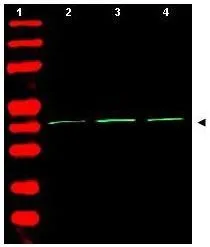
Western Blot of Rabbit Anti-Beta Actin Antibody (GTX21801). Lane 1: molecular weight. Lane 2: human embryonic kidney 293. Lane 3: human lung carcinoma A549. Lane 4: mouse brain. Load: 35 microg per lane. Primary antibody: Beta Actin antibody at 1:1,500 for overnight at 4oC. Secondary antibody: infrared rabbit secondary antibody at 1:10,000 for 45 min at RT. Block: 5% BLOTTO overnight at 4oC. Predicted/Observed size: ~42 kDa corresponding to beta Actin (arrowhead). Other band(s): none


Western Blot of Rabbit Anti-Beta Actin Antibody (GTX21801). Lane 1: molecular weight. Lane 2: human embryonic kidney 293. Lane 3: human lung carcinoma A549. Lane 4: mouse brain. Load: 35 microg per lane. Primary antibody: Beta Actin antibody at 1:1,500 for overnight at 4oC. Secondary antibody: infrared rabbit secondary antibody at 1:10,000 for 45 min at RT. Block: 5% BLOTTO overnight at 4oC. Predicted/Observed size: ~42 kDa corresponding to beta Actin (arrowhead). Other band(s): none
beta Actin antibody
GTX21801
ApplicationsImmunoFluorescence, Western Blot, ELISA, ImmunoCytoChemistry, ImmunoHistoChemistry, ImmunoHistoChemistry Paraffin
Product group Antibodies
TargetACTB
Overview
- SupplierGeneTex
- Product Namebeta Actin antibody
- Delivery Days Customer9
- Application Supplier NoteWB: 1:1000-1:4000. ICC/IF: 1:500-1:2000. ELISA: 1:10000-1:40000. *Optimal dilutions/concentrations should be determined by the researcher.Not tested in other applications.
- ApplicationsImmunoFluorescence, Western Blot, ELISA, ImmunoCytoChemistry, ImmunoHistoChemistry, ImmunoHistoChemistry Paraffin
- CertificationResearch Use Only
- ClonalityPolyclonal
- Concentration1 mg/ml
- ConjugateUnconjugated
- Gene ID60
- Target nameACTB
- Target descriptionactin beta
- Target synonymsBKRNS, BNS, BRWS1, CSMH, DDS1, PS1TP5BP1, THC8, actin, cytoplasmic 1, I(2)-actin, PS1TP5-binding protein 1, beta cytoskeletal actin
- HostRabbit
- IsotypeIgG
- Protein IDP60709
- Protein NameActin, cytoplasmic 1
- Scientific DescriptionThe two major cytoskeletal proteins implicated in cell motility are actin and myosin. Actin and myosin are constituents of many cell types and are involved in a myriad of cellular processes including locomotion, secretion, cytoplasmic streaming, phagocytosis and cytokinesis. Although actin is one of the most conserved eukaryotic proteins, it is expressed in mammals and birds as at least six isoforms characterized by electrophoresis and amino acid sequence analysis. Four of them represent the differentiation markers of muscle tissues and two are found in practically all cells.
- Storage Instruction-20°C or -80°C,2°C to 8°C
- UNSPSC12352203
References
- Up-regulation of histamine H4 receptors contributes to splenic apoptosis in septic mice: counteraction of the antiapoptotic action of nuclear factor-kappaB. Matsuda N et al., 2010 Mar, J Pharmacol Exp TherRead more
Datasheet
Related products
![F-Actin antibody [NH3] detects F-Actin protein at cytoskeleton by immunofluorescent analysis. Sample: Cultured rat E18 primary cortical neuron, DIV 8. Cells were fixed in 4% paraformaldehyde at RT for 15 min. Red: F-Actin protein stained by F-Actin antibody [NH3] (GTX20205) diluted at 1:250. Green: NF-M, stained by NF-M antibody (GTX85461) diluted at 1:250. Blue: Fluoroshield with DAPI (GTX30920).](https://www.genetex.com/upload/website/prouct_img/normal/GTX20205/GTX20205__20161004_IFA_w_23060620_620.webp)
Product group Antibodies
References
F-Actin antibody [NH3]GTX20205
ApplicationsFlow Cytometry, ImmunoFluorescence, Western Blot, ELISA, ImmunoCytoChemistry, ImmunoHistoChemistry, ImmunoHistoChemistry Frozen
TargetACTB
- SizePrice
![ICC/IF analysis of FS-11 cells using GTX26276 beta Actin antibody [AC-15].](https://www.genetex.com/upload/website/prouct_img/normal/GTX26276/GTX26276_20170605_ICCIF_1_w_23060722_508.webp)
Product group Antibodies
References
beta Actin antibody [AC-15]GTX26276
ApplicationsImmunoFluorescence, ImmunoPrecipitation, Western Blot, ELISA, ImmunoCytoChemistry, ImmunoHistoChemistry, ImmunoHistoChemistry Paraffin
TargetACTB
- SizePrice
Product group Antibodies
References
beta Actin antibodyGTX30632
ApplicationsWestern Blot, ImmunoHistoChemistry, ImmunoHistoChemistry Paraffin
TargetACTB
- SizePrice
Product group Antibodies
beta Actin antibodyGTX31732
ApplicationsImmunoFluorescence, Western Blot, ELISA, ImmunoCytoChemistry
TargetACTB
- SizePrice
Product group Antibodies
References
beta Actin antibodyGTX109639
ApplicationsImmunoFluorescence, ImmunoPrecipitation, Western Blot, ImmunoCytoChemistry, ImmunoHistoChemistry, ImmunoHistoChemistry Frozen, ImmunoHistoChemistry Paraffin
TargetACTB
- SizePrice
Product group Antibodies
References
beta Actin antibodyGTX110564
ApplicationsImmunoFluorescence, ImmunoPrecipitation, Western Blot, ImmunoCytoChemistry, ImmunoHistoChemistry, ImmunoHistoChemistry Paraffin
TargetACTB
- SizePrice
Product group Antibodies
References
beta Actin antibody [GT5512]GTX629630
ApplicationsImmunoFluorescence, ImmunoPrecipitation, Western Blot, ImmunoCytoChemistry, ImmunoHistoChemistry, ImmunoHistoChemistry Paraffin
TargetACTB
- SizePrice
Product group Antibodies
beta Actin antibody [HL1926]GTX637675
ApplicationsImmunoFluorescence, Western Blot, ImmunoCytoChemistry, ImmunoHistoChemistry, ImmunoHistoChemistry Paraffin
TargetACTB
- SizePrice
Product group Antibodies
beta Actin antibody [HL1927]GTX637676
ApplicationsWestern Blot, ImmunoHistoChemistry, ImmunoHistoChemistry Paraffin
TargetACTB
- SizePrice
![FACS analysis of MCF-7 cells using GTX83163 beta Actin antibody [8H10D10]. Right : beta Actin Left : negative control](https://www.genetex.com/upload/website/prouct_img/normal/GTX83163/GTX83163_20170912_FACS_w_23061322_375.webp)
Product group Antibodies
References
beta Actin antibody [8H10D10]GTX83163
ApplicationsFlow Cytometry, ImmunoFluorescence, Western Blot, ELISA, ImmunoCytoChemistry
TargetACTB
- SizePrice