

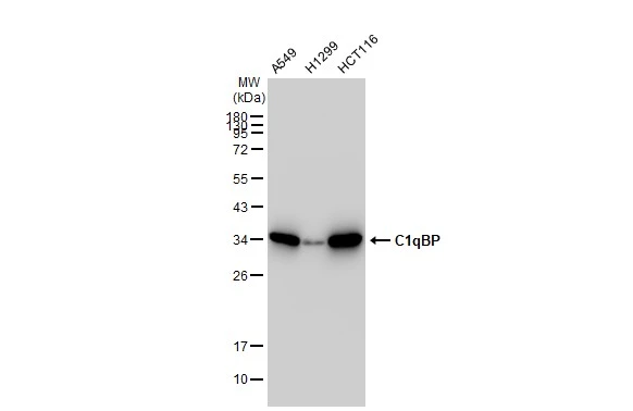
Various whole cell extracts (30 μg) were separated by 12% SDS-PAGE, and the membrane was blotted with C1qBP antibody [GT1250] (GTX02847) diluted at 1:1000. The HRP-conjugated anti-rabbit IgG antibody (GTX213110-01) was used to detect the primary antibody.
![IHC-P analysis of mouse spleen tissue section using GTX02847 C1qBP antibody [GT1250]. Dilution : 1:100](https://www.genetex.com/upload/website/prouct_img/normal/GTX02847/A11292_IHC_03_(1069367)_w_23053123_960.webp "IHC-P analysis of mouse spleen tissue section using GTX02847 C1qBP antibody [GT1250]. Dilution : 1:100")
![Various whole cell extracts (30 μg) were separated by 12% SDS-PAGE, and the membrane was blotted with C1qBP antibody [GT1250] (GTX02847) diluted at 1:1000. The HRP-conjugated anti-rabbit IgG antibody (GTX213110-01) was used to detect the primary antibody.](https://www.genetex.com/upload/website/prouct_img/normal/GTX02847/GTX02847_4000000569_20210122_WB_w_23053123_975.webp "Various whole cell extracts (30 μg) were separated by 12% SDS-PAGE, and the membrane was blotted with C1qBP antibody [GT1250] (GTX02847) diluted at 1:1000. The HRP-conjugated anti-rabbit IgG antibody (GTX213110-01) was used to detect the primary antibody.")
![IHC-P analysis of rat kidney tissue section using GTX02847 C1qBP antibody [GT1250]. Dilution : 1:100](https://www.genetex.com/upload/website/prouct_img/normal/GTX02847/A11292_IHC_01_(1069365)_w_23053123_539.webp "IHC-P analysis of rat kidney tissue section using GTX02847 C1qBP antibody [GT1250]. Dilution : 1:100")
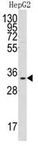
![WB analysis of various samples using GTX02847 C1qBP antibody [GT1250]. Dilution : 1:1000 Loading : 25μg](https://www.genetex.com/upload/website/prouct_img/normal/GTX02847/CutImage_A11292_WB_01_(1069535)_w_23053123_263.webp "WB analysis of various samples using GTX02847 C1qBP antibody [GT1250]. Dilution : 1:1000 Loading : 25μg")
![IHC-P analysis of human colon carcinoma section using GTX02847 C1qBP antibody [GT1250]. Dilution : 1:100](https://www.genetex.com/upload/website/prouct_img/normal/GTX02847/A11292_IHC_02_(1069366)_w_23053123_530.webp "IHC-P analysis of human colon carcinoma section using GTX02847 C1qBP antibody [GT1250]. Dilution : 1:100")
Various whole cell extracts (30 μg) were separated by 12% SDS-PAGE, and the membrane was blotted with C1qBP antibody [GT1250] (GTX02847) diluted at 1:1000. The HRP-conjugated anti-rabbit IgG antibody (GTX213110-01) was used to detect the primary antibody.
C1qBP antibody [GT1250]
GTX02847
ApplicationsWestern Blot, ImmunoHistoChemistry, ImmunoHistoChemistry Paraffin
Product group Antibodies
ReactivityHuman, Mouse, Rat
TargetC1QBP
Overview
- SupplierGeneTex
- Product NameC1qBP antibody [GT1250]
- Delivery Days Customer9
- Application Supplier NoteWB: 1:500 - 1:2000. IHC-P: 1:50 - 1:200. *Optimal dilutions/concentrations should be determined by the researcher.Not tested in other applications.
- ApplicationsWestern Blot, ImmunoHistoChemistry, ImmunoHistoChemistry Paraffin
- CertificationResearch Use Only
- ClonalityMonoclonal
- Clone IDGT1250
- Concentration0.2 mg/ml
- ConjugateUnconjugated
- Gene ID708
- Target nameC1QBP
- Target descriptioncomplement C1q binding protein
- Target synonymsCOXPD33, GC1QBP, HABP1, SF2AP32, SF2p32, gC1Q-R, gC1qR, p32, complement component 1 Q subcomponent-binding protein, mitochondrial, ASF/SF2-associated protein p32, C1q globular domain-binding protein, complement component 1, q subcomponent binding protein, glycoprotein gC1qBP, hyaluronan-binding protein 1, mitochondrial matrix protein p32, p33, splicing factor SF2-associated protein
- HostRabbit
- IsotypeIgG
- Protein IDQ07021
- Protein NameComplement component 1 Q subcomponent-binding protein, mitochondrial
- Scientific DescriptionThe human complement subcomponent C1q associates with C1r and C1s in order to yield the first component of the serum complement system. The protein encoded by this gene is known to bind to the globular heads of C1q molecules and inhibit C1 activation. This protein has also been identified as the p32 subunit of pre-mRNA splicing factor SF2, as well as a hyaluronic acid-binding protein. [provided by RefSeq, Jul 2008]
- ReactivityHuman, Mouse, Rat
- Storage Instruction-20°C,2°C to 8°C
- UNSPSC41116161
Datasheet
Related products

Product group Antibodies
Anti-C1QBP AntibodyA95923
ApplicationsWestern Blot, ELISA, ImmunoHistoChemistry
ReactivityHuman, Mouse, Rat
- SizePrice
Product group Antibodies
Anti-C1QBP Antibody144-01883
ApplicationsImmunoFluorescence, Western Blot
ReactivityHuman, Mouse
TargetC1QBP
- SizePrice

Product group Antibodies
GC1q R Recombinant Antibody, Biotin ConjugatedBSM-61312R-BIOTIN
ApplicationsWestern Blot, ImmunoHistoChemistry, ImmunoHistoChemistry Frozen, ImmunoHistoChemistry Paraffin
TargetC1QBP
- SizePrice

Product group Antibodies
C1QBP AntibodyCSB-PA001089
ApplicationsWestern Blot, ELISA, ImmunoHistoChemistry
ReactivityHuman, Mouse, Rat
TargetC1QBP
- SizePrice
Product group Antibodies
ApplicationsFlow Cytometry
TargetC1QBP
- SizePrice
Product group Antibodies

GC1qR / C1QBP AntibodyLS-C331747
ApplicationsImmunoFluorescence, Western Blot, ImmunoHistoChemistry
ReactivityHuman, Mouse
TargetC1QBP
- SizePrice
Product group Antibodies
C1qBP antibody, InternalGTX81280
ApplicationsWestern Blot
ReactivityHuman
TargetC1QBP
- SizePrice

Product group Antibodies
Anti-C1QBP AntibodyHPA026483
ApplicationsWestern Blot, ImmunoCytoChemistry, ImmunoHistoChemistry
ReactivityHuman, Mouse, Rat
TargetC1QBP
- SizePrice