

C3 antibody
GTX72994
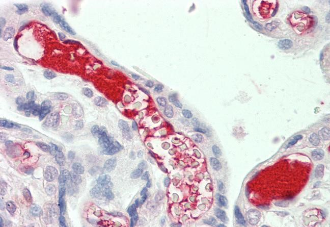
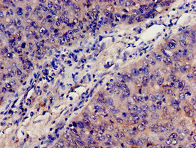
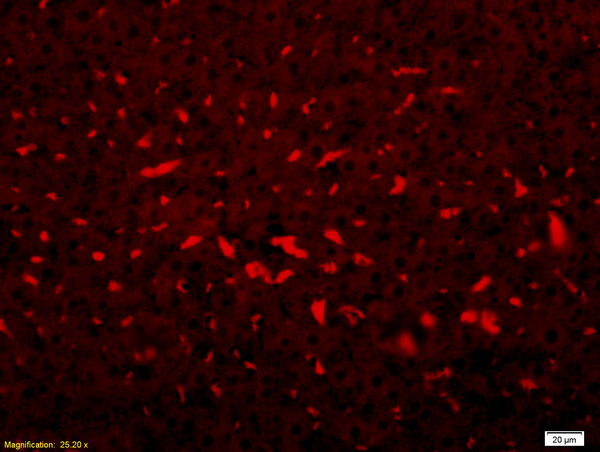
ApplicationsFlow Cytometry, ImmunoElectrophoresis, ImmunoFluorescence, Western Blot, ImmunoCytoChemistry, ImmunoHistoChemistry, ImmunoHistoChemistry Paraffin
Product group Antibodies
ReactivityEquine, Guinea Pig, Hamster, Human, Mouse, Porcine, Primate, Rabbit, Rat
TargetC3
Overview
- SupplierGeneTex
- Product NameC3 antibody
- Delivery Days Customer9
- Application Supplier NoteFor FACS: 1/100. For ICC: 1/500 For Ie: Use at an assay dependent dilution. For WB: 1/1000. Optimal dilutions/concentrations should be determined by the researcher.
- ApplicationsFlow Cytometry, ImmunoElectrophoresis, ImmunoFluorescence, Western Blot, ImmunoCytoChemistry, ImmunoHistoChemistry, ImmunoHistoChemistry Paraffin
- CertificationResearch Use Only
- ClonalityPolyclonal
- ConjugateUnconjugated
- Gene ID718
- Target nameC3
- Target descriptioncomplement C3
- Target synonymsAHUS5, ARMD9, ASP, C3a, C3b, CPAMD1, HEL-S-62p, complement C3, C3 and PZP-like alpha-2-macroglobulin domain-containing protein 1, C3a anaphylatoxin, acylation-stimulating protein cleavage product, complement component 3, complement component C3a, complement component C3b, epididymis secretory sperm binding protein Li 62p, prepro-C3
- HostGoat
- IsotypeIgG
- Protein IDP01024
- Protein NameComplement C3
- Scientific DescriptionComplement component C3 plays a central role in the activation of complement system. Its activation is required for both classical and alternative complement activation pathways. The encoded preproprotein is proteolytically processed to generate alpha and beta subunits that form the mature protein, which is then further processed to generate numerous peptide products. The C3a peptide, also known as the C3a anaphylatoxin, modulates inflammation and possesses antimicrobial activity. Mutations in this gene are associated with atypical hemolytic uremic syndrome and age-related macular degeneration in human patients. [provided by RefSeq, Nov 2015]
- ReactivityEquine, Guinea Pig, Hamster, Human, Mouse, Porcine, Primate, Rabbit, Rat
- Storage Instruction-20°C or -80°C,2°C to 8°C
- UNSPSC41116161
Datasheet
Related products
Product group Antibodies
Anti-C3 AntibodyA286077
ApplicationsELISA, ImmunoHistoChemistry
ReactivityHuman
- SizePrice
Product group Antibodies

Anti-C3b-iC3b [7C12]Ab01554-1.1
ApplicationsFlow Cytometry, ImmunoFluorescence, Western Blot, ELISA
ReactivityHuman
TargetC3
- SizePrice

Product group Antibodies
Anti-C3 Antibody Picoband(r)A00168-3-CARRIER-FREE
ApplicationsFlow Cytometry, Western Blot, ELISA
ReactivityHuman, Mouse, Rat
TargetC3
- SizePrice
Product group Antibodies
C3 AntibodyCSB-PA10599A0RB
ApplicationsImmunoFluorescence, ELISA, ImmunoHistoChemistry
ReactivityHuman
TargetC3
- SizePrice
Product group Antibodies

ApplicationsELISA
ReactivityHuman
TargetC3
- SizePrice
Product group Antibodies
C3 Polyclonal AntibodyCAC09129
ApplicationsImmunoFluorescence, ELISA, ImmunoHistoChemistry
TargetC3
- SizePrice
Product group Antibodies
References
ApplicationsImmunoFluorescence, Western Blot, ELISA, ImmunoHistoChemistry, ImmunoHistoChemistry Frozen, ImmunoHistoChemistry Paraffin
ReactivityHuman
TargetC3
- SizePrice
![The two groups of complement C3 proteins can be purified by GTX02807 C3 antibody [M68] immunoaffinity chromatography from the partially purified milk fractions. Human milk proteins were loaded onto CM-Sepharose 4B column and proteins, including C3, were eluted by different salt (NaCl) concentration. The slat concentration was smaller than 0.3 N. The CM low salt fractions were collected and loaded to the mAb M68-Sepharose column to purify C3. After washing the immunoaffinity column, the captured proteins were eluted by 0.1 N glycine buffer pH 2.4. The eluates were collected into several fractions, E1, E2, E3, E4, E5 and E6. E1-E4 fractions were analyzed by SDS-PAGE and the eluted proteins were stained by CBB. The protein band 2 and 7 as indicated are sliced out and subjected to MS/MS protein identification (by Prottech Inc.), confirming these bands to be complement C3. The relative abundance of peptides matching to C3 is 98% for band 2 and 96% for band 7. For the best detection sensitivity, the samples should be treated under non-boiled and non-reducing conditions. Lane A : Input (partially purified milk fractions) Lane B : Flow through Lane C : Elution 1 Lane D : Elution 2 Lane E : Elution 3 Lane F : Elution 4 Loading : 20 μl](https://www.genetex.com/upload/website/prouct_img/normal/GTX02807/GTX02807_20201130_IP_w_23053122_193.webp)
Product group Antibodies
C3 antibody [M68]GTX02807
ApplicationsImmunoPrecipitation, Western Blot
ReactivityHuman
TargetC3
- SizePrice