

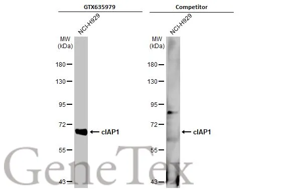
Whole cell extract (30 μg) was separated by 7.5% SDS-PAGE, and the membranes were blotted with cIAP1 antibody [HL1045] (GTX635979) diluted at 1:1000 and competitor's antibody (Competitor) diluted at 1:100. The HRP-conjugated anti-rabbit IgG antibody (GTX213110-01) was used to detect the primary antibody. *The competitor is not affiliated with GeneTex and does not endorse this product.
![cIAP1 antibody [HL1045] detects cIAP1 protein by immunohistochemical analysis. Sample: Paraffin-embedded mouse tissues. cIAP1 stained by cIAP1 antibody [HL1045] (GTX635979) diluted at 1:100. Antigen Retrieval: Citrate buffer, pH 6.0, 15 min](https://www.genetex.com/upload/website/prouct_img/normal/GTX635979/GTX635979_44235_20221227_IHC-P_multiple_M_22122821_390.webp "cIAP1 antibody [HL1045] detects cIAP1 protein by immunohistochemical analysis. Sample: Paraffin-embedded mouse tissues. cIAP1 stained by cIAP1 antibody [HL1045] (GTX635979) diluted at 1:100. Antigen Retrieval: Citrate buffer, pH 6.0, 15 min")
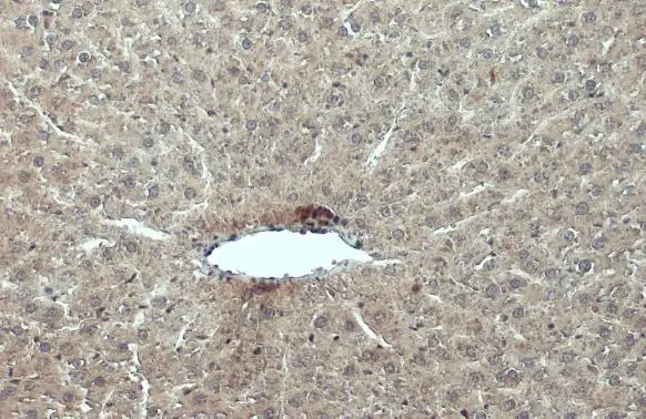
![cIAP1 antibody [HL1045] detects cIAP1 protein at nucleus by immunohistochemical analysis. Sample: Paraffin-embedded dog pancreas. cIAP1 stained by cIAP1 antibody [HL1045] (GTX635979) diluted at 1:100. Antigen Retrieval: Citrate buffer, pH 6.0, 15 min](https://www.genetex.com/upload/website/prouct_img/normal/GTX635979/GTX635979_44235_20230203_IHC-P_Dog_23020621_511.webp "cIAP1 antibody [HL1045] detects cIAP1 protein at nucleus by immunohistochemical analysis. Sample: Paraffin-embedded dog pancreas. cIAP1 stained by cIAP1 antibody [HL1045] (GTX635979) diluted at 1:100. Antigen Retrieval: Citrate buffer, pH 6.0, 15 min")
![cIAP1 antibody [HL1045] detects cIAP1 protein at nucleus by immunohistochemical analysis. Sample: Paraffin-embedded cat lung. cIAP1 stained by cIAP1 antibody [HL1045] (GTX635979) diluted at 1:100. Antigen Retrieval: Citrate buffer, pH 6.0, 15 min](https://www.genetex.com/upload/website/prouct_img/normal/GTX635979/GTX635979_44235_20230203_IHC-P_Cat_23020621_927.webp "cIAP1 antibody [HL1045] detects cIAP1 protein at nucleus by immunohistochemical analysis. Sample: Paraffin-embedded cat lung. cIAP1 stained by cIAP1 antibody [HL1045] (GTX635979) diluted at 1:100. Antigen Retrieval: Citrate buffer, pH 6.0, 15 min")
![Non-transfected (–) and transfected (+) 293T whole cell extracts (50 μg) were separated by 7.5% SDS-PAGE, and the membrane was blotted with cIAP1 antibody [HL1045] (GTX635979) diluted at 1:1000. The HRP-conjugated anti-rabbit IgG antibody (GTX213110-01) was used to detect the primary antibody.](https://www.genetex.com/upload/website/prouct_img/normal/GTX635979/GTX635979_44235_20220128_WB_shRNA_watermark_w_23061202_359.webp "Non-transfected (–) and transfected (+) 293T whole cell extracts (50 μg) were separated by 7.5% SDS-PAGE, and the membrane was blotted with cIAP1 antibody [HL1045] (GTX635979) diluted at 1:1000. The HRP-conjugated anti-rabbit IgG antibody (GTX213110-01) was used to detect the primary antibody.")
![cIAP1 antibody [HL1045] detects cIAP1 protein at cytoplasm and nucleus by immunohistochemical analysis. Sample: Paraffin-embedded rat spleen. cIAP1 stained by cIAP1 antibody [HL1045] (GTX635979) diluted at 1:100. Antigen Retrieval: Citrate buffer, pH 6.0, 15 min](https://www.genetex.com/upload/website/prouct_img/normal/GTX635979/GTX635979_44235_20220311_IHC-P_R_w_23061202_340.webp "cIAP1 antibody [HL1045] detects cIAP1 protein at cytoplasm and nucleus by immunohistochemical analysis. Sample: Paraffin-embedded rat spleen. cIAP1 stained by cIAP1 antibody [HL1045] (GTX635979) diluted at 1:100. Antigen Retrieval: Citrate buffer, pH 6.0, 15 min")
![Non-transfected (–) and transfected (+) 293T whole cell extracts (30 μg) were separated by 7.5% SDS-PAGE, and the membrane was blotted with cIAP1 antibody [HL1045] (GTX635979) diluted at 1:5000. The HRP-conjugated anti-rabbit IgG antibody (GTX213110-01) was used to detect the primary antibody.](https://www.genetex.com/upload/website/prouct_img/normal/GTX635979/GTX635979_44235_20210827_WB_B_w_23061202_846.webp "Non-transfected (–) and transfected (+) 293T whole cell extracts (30 μg) were separated by 7.5% SDS-PAGE, and the membrane was blotted with cIAP1 antibody [HL1045] (GTX635979) diluted at 1:5000. The HRP-conjugated anti-rabbit IgG antibody (GTX213110-01) was used to detect the primary antibody.")
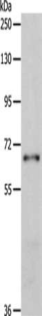
![Whole cell extract (30 μg) was separated by 7.5% SDS-PAGE, and the membrane was blotted with cIAP1 antibody [HL1045] (GTX635979) diluted at 1:1000. The HRP-conjugated anti-rabbit IgG antibody (GTX213110-01) was used to detect the primary antibody.](https://www.genetex.com/upload/website/prouct_img/normal/GTX635979/GTX635979_44235_20210305_WB_w_23061202_471.webp "Whole cell extract (30 μg) was separated by 7.5% SDS-PAGE, and the membrane was blotted with cIAP1 antibody [HL1045] (GTX635979) diluted at 1:1000. The HRP-conjugated anti-rabbit IgG antibody (GTX213110-01) was used to detect the primary antibody.")
Whole cell extract (30 μg) was separated by 7.5% SDS-PAGE, and the membranes were blotted with cIAP1 antibody [HL1045] (GTX635979) diluted at 1:1000 and competitor's antibody (Competitor) diluted at 1:100. The HRP-conjugated anti-rabbit IgG antibody (GTX213110-01) was used to detect the primary antibody. *The competitor is not affiliated with GeneTex and does not endorse this product.
cIAP1 antibody [HL1045]
GTX635979
ApplicationsWestern Blot, ImmunoHistoChemistry, ImmunoHistoChemistry Paraffin
Product group Antibodies
ReactivityCanine, Feline, Human, Mouse, Rat
TargetBIRC2
Overview
- SupplierGeneTex
- Product NamecIAP1 antibody [HL1045]
- Delivery Days Customer9
- Application Supplier NoteWB: 1:500-1:3000. *Optimal dilutions/concentrations should be determined by the researcher.Not tested in other applications.
- ApplicationsWestern Blot, ImmunoHistoChemistry, ImmunoHistoChemistry Paraffin
- CertificationResearch Use Only
- ClonalityMonoclonal
- Clone IDHL1045
- Concentration1 mg/ml
- ConjugateUnconjugated
- Gene ID329
- Target nameBIRC2
- Target descriptionbaculoviral IAP repeat containing 2
- Target synonymsAPI1, HIAP2, Hiap-2, MIHB, RNF48, c-IAP1, cIAP1, baculoviral IAP repeat-containing protein 2, IAP homolog B, IAP-2, NFR2-TRAF signalling complex protein, RING finger protein 48, RING-type E3 ubiquitin transferase BIRC2, TNFR2-TRAF-signaling complex protein 2, apoptosis inhibitor 1, cellular inhibitor of apoptosis 1, inhibitor of apoptosis protein 2
- HostRabbit
- IsotypeIgG
- Protein IDQ13490
- Protein NameBaculoviral IAP repeat-containing protein 2
- Scientific DescriptionThe protein encoded by this gene is a member of a family of proteins that inhibits apoptosis by binding to tumor necrosis factor receptor-associated factors TRAF1 and TRAF2, probably by interfering with activation of ICE-like proteases. This encoded protein inhibits apoptosis induced by serum deprivation and menadione, a potent inducer of free radicals. Alternatively spliced transcript variants encoding different isoforms have been found for this gene. [provided by RefSeq, Jan 2012]
- ReactivityCanine, Feline, Human, Mouse, Rat
- Storage Instruction-20°C or -80°C,2°C to 8°C
- UNSPSC41116161
Datasheet
Related products
Product group Antibodies
BIRC2 AntibodyCSB-PA191509
ApplicationsWestern Blot, ELISA
ReactivityHuman, Mouse
TargetBIRC2
- SizePrice

Product group Antibodies
Anti-cIAP1/BIRC2 Antibody Picoband(r)A01700-1-CARRIER-FREE
ApplicationsFlow Cytometry, ImmunoFluorescence, Western Blot, ELISA, ImmunoCytoChemistry
ReactivityHuman, Mouse, Rat
TargetBIRC2
- SizePrice
Product group Antibodies
Anti-BIRC2 AntibodyA28642
ApplicationsWestern Blot
ReactivityHuman, Mouse, Rat
- SizePrice
Product group Antibodies

BIRC2 / cIAP1 Antibody (aa1-300)LS-C746764
ApplicationsImmunoFluorescence, Western Blot
ReactivityHuman, Mouse, Rat
TargetBIRC2
- SizePrice

Product group Antibodies
Anti-BIRC2 AntibodyHPA005513
ApplicationsWestern Blot, ImmunoHistoChemistry
ReactivityHuman, Rat
TargetBIRC2
- SizePrice
Product group Antibodies
BIRC2 Polyclonal AntibodyCAC14894
ApplicationsWestern Blot, ELISA
ReactivityMouse, Rat
TargetBIRC2
- SizePrice
Product group Antibodies
cIAP1 antibodyGTX110087
ApplicationsWestern Blot, ImmunoHistoChemistry, ImmunoHistoChemistry Paraffin
ReactivityHuman, Mouse, Rat
TargetBIRC2
- SizePrice
![Non-transfected (–) and transfected (+) 293T whole cell extracts (30 μg) were separated by 7.5% SDS-PAGE, and the membrane was blotted with cIAP1 antibody [GT422] (GTX629402) diluted at 1:1000. The HRP-conjugated anti-mouse IgG antibody (GTX213111-01) was used to detect the primary antibody.](https://www.genetex.com/upload/website/prouct_img/normal/GTX629402/GTX629402_41344_20180914_WB_B_w_23061202_916.webp)
Product group Antibodies
cIAP1 antibody [GT422]GTX629402
ApplicationsImmunoFluorescence, Western Blot, ImmunoCytoChemistry
ReactivityHuman
TargetBIRC2
- SizePrice