

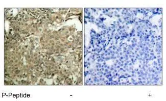
IHC-P image using GTX12866 - Detection of Cofilin by IHC-P in human breast carcinoma tissue.
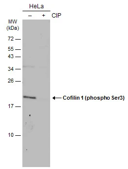
 and cofilin (phospho-Ser3) antibody (GTX12866, Lane 3 and 4)")
IHC-P image using GTX12866 - Detection of Cofilin by IHC-P in human breast carcinoma tissue.
Cofilin 1 (phospho Ser3) antibody
GTX12866
ApplicationsWestern Blot, ImmunoHistoChemistry, ImmunoHistoChemistry Paraffin
Product group Antibodies
ReactivityHuman, Mouse
TargetCFL1
Overview
- SupplierGeneTex
- Product NameCofilin 1 (phospho Ser3) antibody
- Delivery Days Customer9
- ApplicationsWestern Blot, ImmunoHistoChemistry, ImmunoHistoChemistry Paraffin
- CertificationResearch Use Only
- ClonalityPolyclonal
- ConjugateUnconjugated
- Gene ID1072
- Target nameCFL1
- Target descriptioncofilin 1
- Target synonymsCFL, HEL-S-15, cofilin, cofilin-1, 18 kDa phosphoprotein, cofilin 1 (non-muscle), epididymis secretory protein Li 15, p18
- HostRabbit
- IsotypeIgG
- Protein IDP23528
- Protein NameCofilin-1
- Scientific DescriptionThe protein encoded by this gene can polymerize and depolymerize F-actin and G-actin in a pH-dependent manner. Increased phosphorylation of this protein by LIM kinase aids in Rho-induced reorganization of the actin cytoskeleton. Cofilin is a widely distributed intracellular actin-modulating protein that binds and depolymerizes filamentous F-actin and inhibits the polymerization of monomeric G-actin in a pH-dependent manner. It is involved in the translocation of actin-cofilin complex from cytoplasm to nucleus.[supplied by OMIM, Apr 2004]
- ReactivityHuman, Mouse
- Storage Instruction-20°C or -80°C,2°C to 8°C
- UNSPSC12352203
References
- Toyo-oka K, Wachi T, Hunt RF, et al. 14-3-3ε and ζ regulate neurogenesis and differentiation of neuronal progenitor cells in the developing brain. J Neurosci. 2014,34(36):12168-81. doi: 10.1523/JNEUROSCI.2513-13.2014Read this paper
Datasheet
Related products

Product group Antibodies
Anti-CFL1 Antibody144-01704
ApplicationsImmunoFluorescence, Western Blot, ImmunoHistoChemistry
ReactivityHuman, Mouse, Rat
TargetCFL1
- SizePrice
Product group Antibodies
Cofilin 1 (phospho Ser3) antibodyGTX133865
ApplicationsWestern Blot
ReactivityHuman, Mouse
TargetCFL1
- SizePrice
![Cofilin 1 antibody detects Cofilin 1 protein at cytoplasm and nucleus by immunofluorescent analysis. Samples: HeLa cells mock (left) and treated with 0.012 N NaOH/PBS for 1 min (right) were fixed in 4% paraformaldehyde at RT for 15 min. Green: Cofilin 1 protein stained by Cofilin 1 antibody (GTX102156) diluted at 1:1000. Red: alpha Tubulin, a cytoskeleton marker, stained by alpha Tubulin antibody [GT114] (GTX628802) diluted at 1:1000.](https://www.genetex.com/upload/website/prouct_img/normal/GTX102156/GTX102156_40051_20150410_IFA_w_23060100_118.webp)
Product group Antibodies
References
Cofilin 1 antibodyGTX102156
ApplicationsImmunoFluorescence, Western Blot, ImmunoCytoChemistry, ImmunoHistoChemistry, ImmunoHistoChemistry Frozen, ImmunoHistoChemistry Paraffin
ReactivityHuman, Mouse, Rat
TargetCFL1
- SizePrice
Product group Antibodies
Cofilin 1 antibody [AT1C1]GTX57545
ApplicationsFlow Cytometry, ImmunoFluorescence, Western Blot, ImmunoCytoChemistry
ReactivityHuman
TargetCFL1
- SizePrice
![Various whole cell extracts (30 μg) were separated by 15% SDS-PAGE, and the membrane was blotted with Cofilin 1 antibody [GT567] (GTX628804) diluted at 1:1000. The HRP-conjugated anti-mouse IgG antibody (GTX213111-01) was used to detect the primary antibody. Corresponding RNA expression data for the same cell lines are based on Human Protein Atlas program.](https://www.genetex.com/upload/website/prouct_img/normal/GTX628804/GTX628804_41204_20230210_WB_TPM_watermark_23021401_497.webp)
Product group Antibodies
Cofilin 1 antibody [GT567]GTX628804
ApplicationsImmunoFluorescence, Western Blot, ImmunoCytoChemistry, ImmunoHistoChemistry, ImmunoHistoChemistry Paraffin
ReactivityHuman, Mouse, Rat
TargetCFL1
- SizePrice
![Various whole cell extracts (30 μg) were separated by 12% SDS-PAGE, and the membrane was blotted with Cofilin 1 antibody [GT217] (GTX632582) diluted at 1:1000. The HRP-conjugated anti-mouse IgG antibody (GTX213111-01) was used to detect the primary antibody.](https://www.genetex.com/upload/website/prouct_img/normal/GTX632582/GTX632582_42184_20170615_WB_M_w_23061202_342.webp)
Product group Antibodies
Cofilin 1 antibody [GT217]GTX632582
ApplicationsImmunoFluorescence, ImmunoPrecipitation, Western Blot, ImmunoCytoChemistry, ImmunoHistoChemistry, ImmunoHistoChemistry Paraffin
ReactivityHuman, Mouse, Rat
TargetCFL1
- SizePrice
Product group Antibodies
ApplicationsImmunoPrecipitation, Western Blot, ImmunoCytoChemistry, ImmunoHistoChemistry
ReactivityMouse
TargetCFL1
- SizePrice
Product group Antibodies
References
ApplicationsImmunoFluorescence, Western Blot, ImmunoCytoChemistry, ImmunoHistoChemistry, ImmunoHistoChemistry Frozen, ImmunoHistoChemistry Paraffin
ReactivityBovine, Equine, Human, Mouse, Porcine, Rat, Sheep
TargetCFL1
- SizePrice
Product group Antibodies
CFL1 AntibodyCSB-PA001739
ApplicationsImmunoFluorescence, Western Blot, ELISA, ImmunoHistoChemistry
ReactivityHuman, Mouse, Rat
TargetCFL1
- SizePrice