

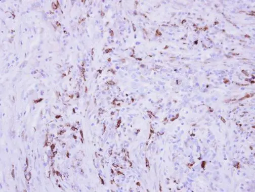
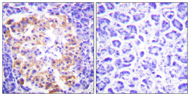
Immunohistochemical analysis of paraffin-embedded human PAPILLARY CA_STROMAL CELLS, using Collagen III (GTX102997) antibody at 1:250 dilution.
Antigen Retrieval: Trilogy? (EDTA based, pH 8.0) buffer, 15min
![Non-transfected (–) and transfected (+) HeLa whole cell extracts (30 μg) were separated by 5% SDS-PAGE, and the membrane was blotted with Collagen III antibody [C2C3], C-term (GTX102997) diluted at 1:2000. The HRP-conjugated anti-rabbit IgG antibody (GTX213110-01) was used to detect the primary antibody.](https://www.genetex.com/upload/website/prouct_img/normal/GTX102997/GTX102997_42796_20181221_WB_shRNA_watermark_23121122_933.webp "Non-transfected (–) and transfected (+) HeLa whole cell extracts (30 μg) were separated by 5% SDS-PAGE, and the membrane was blotted with Collagen III antibody [C2C3], C-term (GTX102997) diluted at 1:2000. The HRP-conjugated anti-rabbit IgG antibody (GTX213110-01) was used to detect the primary antibody.")
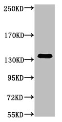
![Various whole cell extracts (30 μg) were separated by 5% SDS-PAGE, and the membrane was blotted with Collagen III antibody [C2C3], C-term (GTX102997) diluted at 1:1000. The HRP-conjugated anti-rabbit IgG antibody (GTX213110-01) was used to detect the primary antibody.](https://www.genetex.com/upload/website/prouct_img/normal/GTX102997/GTX102997_44566_20220121_WB_24110700_835.webp "Various whole cell extracts (30 μg) were separated by 5% SDS-PAGE, and the membrane was blotted with Collagen III antibody [C2C3], C-term (GTX102997) diluted at 1:1000. The HRP-conjugated anti-rabbit IgG antibody (GTX213110-01) was used to detect the primary antibody.")
Immunohistochemical analysis of paraffin-embedded human PAPILLARY CA_STROMAL CELLS, using Collagen III (GTX102997) antibody at 1:250 dilution.
Antigen Retrieval: Trilogy? (EDTA based, pH 8.0) buffer, 15min
Collagen III antibody [C2C3], C-term
GTX102997
ApplicationsImmunoFluorescence, Western Blot, ImmunoCytoChemistry, ImmunoHistoChemistry, ImmunoHistoChemistry Paraffin
Product group Antibodies
ReactivityHuman, Rat
TargetCOL3A1
Overview
- SupplierGeneTex
- Product NameCollagen III antibody [C2C3], C-term
- Delivery Days Customer9
- Application Supplier NoteWB: 1:500-1:3000. IHC-P: 1:100-1:1000. *Optimal dilutions/concentrations should be determined by the researcher.Not tested in other applications.
- ApplicationsImmunoFluorescence, Western Blot, ImmunoCytoChemistry, ImmunoHistoChemistry, ImmunoHistoChemistry Paraffin
- CertificationResearch Use Only
- ClonalityPolyclonal
- Concentration1.32 mg/ml
- ConjugateUnconjugated
- Gene ID1281
- Target nameCOL3A1
- Target descriptioncollagen type III alpha 1 chain
- Target synonymsEDS4A, EDSVASC, PMGEDSV, collagen alpha-1(III) chain, Ehlers-Danlos syndrome type IV, autosomal dominant, alpha-1 type III collagen, alpha1 (III) collagen, collagen, fetal, collagen, type III, alpha 1
- HostRabbit
- IsotypeIgG
- Protein IDP02461
- Protein NameCollagen alpha-1(III) chain
- Scientific DescriptionThis gene encodes the pro-alpha1 chains of type III collagen, a fibrillar collagen that is found in extensible connective tissues such as skin, lung, uterus, intestine and the vascular system, frequently in association with type I collagen. Mutations in this gene are associated with Ehlers-Danlos syndrome types IV, and with aortic and arterial aneurysms. Two transcripts, resulting from the use of alternate polyadenylation signals, have been identified for this gene. [provided by R. Dalgleish]
- ReactivityHuman, Rat
- Storage Instruction-20°C or -80°C,2°C to 8°C
- UNSPSC41116161
Datasheet
Related products
Product group Antibodies
ApplicationsImmunoFluorescence, ELISA, ImmunoHistoChemistry
ReactivityHuman, Mouse, Rat
- SizePrice

Product group Antibodies
Anti-Collagen III/COL3A1 Antibody Picoband(r)A00788-3-CARRIER-FREE
ApplicationsFlow Cytometry, Western Blot, ELISA, ImmunoHistoChemistry
ReactivityHuman, Mouse, Rat
TargetCOL3A1
- SizePrice
Product group Antibodies
Anti-COL3A1 Antibody144-03795
ApplicationsWestern Blot
ReactivityHuman, Mouse
TargetCOL3A1
- SizePrice
Product group Antibodies

ApplicationsImmunoFluorescence, Western Blot, ImmunoHistoChemistry, ImmunoHistoChemistry Paraffin
ReactivityHuman, Mouse, Rat
TargetCOL3A1
- SizePrice

Product group Antibodies
References
Collagen III Polyclonal AntibodyBS-0549R
ApplicationsImmunoFluorescence, Western Blot, ImmunoCytoChemistry, ImmunoHistoChemistry, ImmunoHistoChemistry Frozen, ImmunoHistoChemistry Paraffin
ReactivityHuman
TargetCOL3A1
- SizePrice
Product group Antibodies
COL3A1 Monoclonal AntibodyCSB-MA000282
ApplicationsWestern Blot, ELISA
ReactivityHuman, Mouse, Rat
TargetCOL3A1
- SizePrice
Product group Antibodies

Procollagen Type III, human, bovineCO23311-0.1
ApplicationsImmunoFluorescence, Western Blot, ELISA, ImmunoHistoChemistry, ImmunoHistoChemistry Paraffin, RadioImmunoAssay
ReactivityBovine, Human, Porcine
TargetCOL3A1
- SizePrice
Product group Antibodies
ApplicationsFlow Cytometry
TargetCOL3A1
- SizePrice
![IHC-Fr analysis of rat skin tissue using GTX26310 Collagen III antibody [FH-7A] at 1:8,000.](https://www.genetex.com/upload/website/prouct_img/normal/GTX26310/GTX26310_20170605_IHC-Fr_w_23060722_325.webp)
Product group Antibodies
References
Collagen III antibody [FH-7A]GTX26310
ApplicationsDot Blot, Western Blot, ELISA, ImmunoHistoChemistry, ImmunoHistoChemistry Frozen, ImmunoHistoChemistry Paraffin
ReactivityHuman, Rabbit, Rat
TargetCOL3A1
- SizePrice