

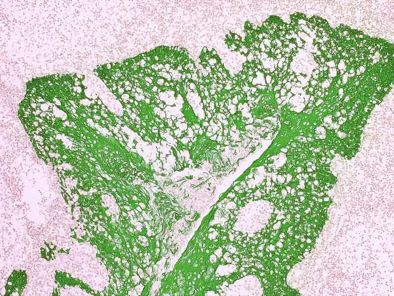
IHC-P analysis of human tonsil tissue using GTX01662 Cytokeratin 7 + 8 antibody [CAM5.2] (ready-to-use) with GTX03612 Tissue Glue.
IHC-P analysis of human tonsil tissue using GTX01662 Cytokeratin 7 + 8 antibody [CAM5.2] (ready-to-use) with GTX03612 Tissue Glue.
Cytokeratin 7 + 8 antibody [CAM5.2] (ready-to-use)
GTX01662
ApplicationsImmunoHistoChemistry, ImmunoHistoChemistry Paraffin
Product group Antibodies
ReactivityHuman
TargetKRT7
Overview
- SupplierGeneTex
- Product NameCytokeratin 7 + 8 antibody [CAM5.2] (ready-to-use)
- Delivery Days Customer9
- Application Supplier NoteIHC-P: . *Optimal dilutions/concentrations should be determined by the researcher.Not tested in other applications.
- ApplicationsImmunoHistoChemistry, ImmunoHistoChemistry Paraffin
- CertificationResearch Use Only
- ClonalityMonoclonal
- Clone IDCAM5.2
- ConjugateUnconjugated
- Gene ID3855
- Target nameKRT7
- Target descriptionkeratin 7
- Target synonymsCK7, K2C7, K7, SCL, keratin, type II cytoskeletal 7, CK-7, cytokeratin 7, keratin 7, type II, keratin, 55K type II cytoskeletal, keratin, simple epithelial type I, K7, sarcolectin, type II mesothelial keratin K7, type-II keratin Kb7
- HostMouse
- IsotypeIgG2a
- Protein IDP05787
- Protein NameKeratin, type II cytoskeletal 8
- ReactivityHuman
- Storage Instruction2°C to 8°C
- UNSPSC12352203
References
- Tamura N, Maeda H, Nishikori M, et al. Correction to: Histologic transformation of t(11;18)-positive MALT lymphoma presented with aberrant T-cell marker expression. Int J Hematol. 2020,112(2):265. doi: 10.1007/s12185-020-02924-8Read this paper
- Tsuzuki S, Kataoka TR, Ito H, et al. A case of renal cell carcinoma unclassified with medullary phenotype without detectable gene deletion. Pathol Int. 2019,69(12):710-714. doi: 10.1111/pin.12858Read this paper
- Yu W, Wang Y, Rao Q, et al. Xp11.2 translocation renal neoplasm with features of TFE3 rearrangement associated renal cell carcinoma and Xp11 translocation renal mesenchymal tumor with melanocytic differentiation harboring NONO-TFE3 fusion gene. Pathol Res Pract. 2019,215(9):152521. doi: 10.1016/j.prp.2019.152521Read this paper
- Asa SL, Ezzat S, Kelly DF, et al. Hypothalamic Vasopressin-Producing Tumors: Often Inappropriate Diuresis But Occasionally Cushing Disease. Am J Surg Pathol. 2019,43(2):251-260. doi: 10.1097/PAS.0000000000001185Read this paper
- Thompson LD, Aslam MN, Stall JN, et al. Clinicopathologic and Immunophenotypic Characterization of 25 Cases of Acinic Cell Carcinoma with High-Grade Transformation. Head Neck Pathol. 2016,10(2):152-60. doi: 10.1007/s12105-015-0645-xRead this paper
Datasheet
Related products

Product group Antibodies
Anti-KRT7 Antibody Picoband(r)A02416-2-CARRIER-FREE
ApplicationsWestern Blot, ImmunoHistoChemistry
ReactivityHuman
TargetKRT7
- SizePrice
Product group Antibodies
KRT7 Polyclonal AntibodyCAC14736
ApplicationsImmunoFluorescence, ImmunoPrecipitation, Western Blot, ELISA, ImmunoHistoChemistry
TargetKRT7
- SizePrice
Product group Antibodies
Anti-Cytokeratin 7 Antibody188-10504
ApplicationsFlow Cytometry
ReactivityHuman
TargetKRT7
- SizePrice
Product group Antibodies
References
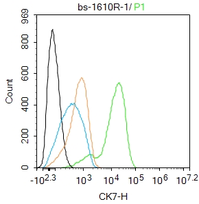
CK7 Polyclonal AntibodyBS-1610R
ApplicationsFlow Cytometry, ImmunoFluorescence, Western Blot, ELISA, ImmunoCytoChemistry, ImmunoHistoChemistry, ImmunoHistoChemistry Frozen, ImmunoHistoChemistry Paraffin
ReactivityHuman, Mouse, Rat
TargetKRT7
- SizePrice
Product group Antibodies
ApplicationsImmunoHistoChemistry, ImmunoHistoChemistry Paraffin
ReactivityHuman
TargetKRT7
- SizePrice
![FACS analysis of HeLa cells using GTX83354 Cytokeratin 7 antibody [5D12]. Green : Cytokeratin 7 Purple : negative control](https://www.genetex.com/upload/website/prouct_img/normal/GTX83354/GTX83354_20170912_FACS_w_23061322_967.webp)
Product group Antibodies
Cytokeratin 7 antibody [5D12]GTX83354
ApplicationsFlow Cytometry, Western Blot, ELISA
ReactivityHuman
TargetKRT7
- SizePrice
Product group Antibodies
References
Cytokeratin 7 antibody [N1C2]GTX109723
ApplicationsImmunoFluorescence, Western Blot, ImmunoCytoChemistry, ImmunoHistoChemistry, ImmunoHistoChemistry Frozen, ImmunoHistoChemistry Paraffin
ReactivityHuman
TargetKRT7
- SizePrice
Product group Antibodies
References
ApplicationsImmunoFluorescence, Western Blot, ImmunoCytoChemistry, ImmunoHistoChemistry, ImmunoHistoChemistry Paraffin
ReactivityHuman, Mouse
TargetKRT7
- SizePrice
Product group Antibodies
ApplicationsImmunoHistoChemistry, ImmunoHistoChemistry Paraffin
ReactivityHuman
TargetKRT7
- SizePrice
![IHC-P analysis of invasive breast carcinoma tissue using GTX01903 Cytokeratin 7 antibody [RN7]. Note the intense staining of invasive tumor cells.](https://www.genetex.com/upload/website/prouct_img/normal/GTX01903/GTX01903_20200811_IHC-P_81_w_23053121_225.webp)
Product group Antibodies
Cytokeratin 7 antibody [RN7]GTX01903
ApplicationsImmunoHistoChemistry, ImmunoHistoChemistry Paraffin
ReactivityHuman
TargetKRT7
- SizePrice