

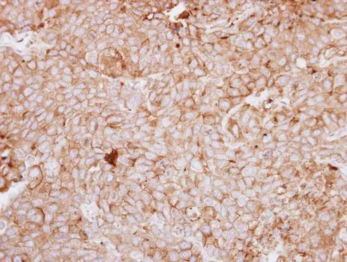
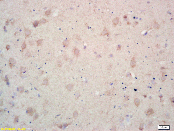
Immunohistochemical analysis of paraffin-embedded BT474 xenograft, using DDR1(GTX111312) antibody at 1:250 dilution.
Antigen Retrieval: Trilogy? (EDTA based, pH 8.0) buffer, 15min
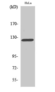
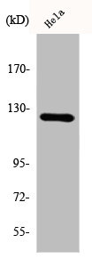
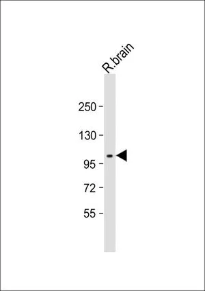
 A: THP-1 7.5% SDS PAGE GTX111312 diluted at 1:500 The HRP-conjugated anti-rabbit IgG antibody (GTX213110-01) was used to detect the primary antibody.")
Immunohistochemical analysis of paraffin-embedded BT474 xenograft, using DDR1(GTX111312) antibody at 1:250 dilution.
Antigen Retrieval: Trilogy? (EDTA based, pH 8.0) buffer, 15min
DDR1 antibody [C1C3]
GTX111312
ApplicationsWestern Blot, ImmunoHistoChemistry, ImmunoHistoChemistry Paraffin
Product group Antibodies
ReactivityHuman
TargetDDR1
Overview
- SupplierGeneTex
- Product NameDDR1 antibody [C1C3]
- Delivery Days Customer9
- Application Supplier NoteWB: 1:500-1:3000. IHC-P: 1:100-1:1000. *Optimal dilutions/concentrations should be determined by the researcher.Not tested in other applications.
- ApplicationsWestern Blot, ImmunoHistoChemistry, ImmunoHistoChemistry Paraffin
- CertificationResearch Use Only
- ClonalityPolyclonal
- Concentration1.03 mg/ml
- ConjugateUnconjugated
- Gene ID780
- Target nameDDR1
- Target descriptiondiscoidin domain receptor tyrosine kinase 1
- Target synonymsCAK, CD167, DDR, EDDR1, HGK2, MCK10, NEP, NTRK4, PTK3, PTK3A, RTK6, TRKE, epithelial discoidin domain-containing receptor 1, CD167 antigen-like family member A, PTK3A protein tyrosine kinase 3A, cell adhesion kinase, mammary carcinoma kinase 10, neuroepithelial tyrosine kinase, neurotrophic tyrosine kinase, receptor, type 4, protein-tyrosine kinase RTK-6, tyrosine kinase DDR, tyrosine-protein kinase CAK
- HostRabbit
- IsotypeIgG
- Protein IDQ08345
- Protein NameEpithelial discoidin domain-containing receptor 1
- Scientific DescriptionReceptor tyrosine kinases (RTKs) play a key role in the communication of cells with their microenvironment. These molecules are involved in the regulation of cell growth, differentiation and metabolism. The protein encoded by this gene is a RTK that is widely expressed in normal and transformed epithelial cells and is activated by various types of collagen. This protein belongs to a subfamily of tyrosine kinase receptors with a homology region to the Dictyostelium discoideum protein discoidin I in their extracellular domain. Its autophosphorylation is achieved by all collagens so far tested (type I to type VI). In situ studies and Northern-blot analysis showed that expression of this encoded protein is restricted to epithelial cells, particularly in the kidney, lung, gastrointestinal tract, and brain. In addition, this protein is significantly over-expressed in several human tumors from breast, ovarian, esophageal, and pediatric brain. This gene is located on chromosome 6p21.3 in proximity to several HLA class I genes. Alternative splicing of this gene results in multiple transcript variants. [provided by RefSeq]
- ReactivityHuman
- Storage Instruction-20°C or -80°C,2°C to 8°C
- UNSPSC41116161
Datasheet
Related products
Product group Antibodies

Anti-Discoidin Domain Receptor 1 [3E3]AB00438-1.1-BT
ApplicationsWestern Blot, ELISA, Neutralisation/Blocking
ReactivityHuman
TargetDDR1
- SizePrice
Product group Antibodies
Anti-DDR1 AntibodyA101481
ApplicationsWestern Blot, ELISA
ReactivityHuman
- SizePrice

Product group Antibodies
Anti-MCK10/NEP/DDR1 Antibody Picoband(r)A00905-CARRIER-FREE
ApplicationsFlow Cytometry, ImmunoFluorescence, Western Blot, ELISA, ImmunoCytoChemistry, ImmunoHistoChemistry
ReactivityHuman, Mouse, Rat
TargetDDR1
- SizePrice
Product group Antibodies
Anti-Phospho-DDR1-Y792 Antibody144-50864
ApplicationsWestern Blot
ReactivityHuman, Mouse, Rat
TargetDDR1
- SizePrice
Product group Antibodies

MCK10 AntibodyABX033584
ApplicationsWestern Blot, ELISA, ImmunoHistoChemistry
- SizePrice
Product group Antibodies

NEP / DDR1 Antibody (Tyr513)LS-C769154
ApplicationsWestern Blot, ELISA
ReactivityHuman, Mouse, Rat
TargetDDR1
- SizePrice
Product group Antibodies
References
DDR1 Polyclonal AntibodyBS-0671R
ApplicationsImmunoFluorescence, Western Blot, ELISA, ImmunoCytoChemistry, ImmunoHistoChemistry, ImmunoHistoChemistry Frozen, ImmunoHistoChemistry Paraffin
ReactivityHuman, Mouse, Rat
TargetDDR1
- SizePrice
Product group Antibodies
DDR1 AntibodyCSB-PA002098
ApplicationsWestern Blot, ELISA
ReactivityHuman
TargetDDR1
- SizePrice
Product group Antibodies
ApplicationsImmunoPrecipitation, Western Blot, ImmunoCytoChemistry, ImmunoHistoChemistry
ReactivityMouse
TargetDDR1
- SizePrice
Product group Antibodies
References
DDR1 antibodyGTX25508
ApplicationsFlow Cytometry, Western Blot, ImmunoHistoChemistry, ImmunoHistoChemistry Paraffin
ReactivityHuman, Rat
TargetDDR1
- SizePrice