

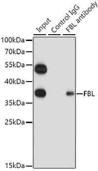
IP analysis of HeLa cell lysate using GTX65859 Fibrillarin antibody. Antibody amount : 1μg / 200μg lysate Dilution : 1:1000


IP analysis of HeLa cell lysate using GTX65859 Fibrillarin antibody. Antibody amount : 1μg / 200μg lysate Dilution : 1:1000
Fibrillarin antibody
GTX65859
ApplicationsImmunoFluorescence, ImmunoPrecipitation, Western Blot, ImmunoCytoChemistry, ImmunoHistoChemistry, ImmunoHistoChemistry Paraffin
Product group Antibodies
ReactivityHuman, Monkey, Mouse, Rat
TargetFBL
Overview
- SupplierGeneTex
- Product NameFibrillarin antibody
- Delivery Days Customer9
- Application Supplier NoteWB: 1:500 - 1:2000. ICC/IF: 1:50 - 1:200. IHC-P: 1:50 - 1:200. IP: 1:20 - 1:50. *Optimal dilutions/concentrations should be determined by the researcher.Not tested in other applications.
- ApplicationsImmunoFluorescence, ImmunoPrecipitation, Western Blot, ImmunoCytoChemistry, ImmunoHistoChemistry, ImmunoHistoChemistry Paraffin
- CertificationResearch Use Only
- ClonalityPolyclonal
- ConjugateUnconjugated
- Gene ID2091
- Target nameFBL
- Target descriptionfibrillarin
- Target synonymsFIB, FLRN, Nop1, RNU3IP1, rRNA 2'-O-methyltransferase fibrillarin, 34 kDa nucleolar scleroderma antigen, 34-kD nucleolar scleroderma antigen, RNA, U3 small nucleolar interacting protein 1, U6 snRNA 2'-O-methyltransferase fibrillarin, histone-glutamine methyltransferase
- HostRabbit
- IsotypeIgG
- Protein IDP22087
- Protein NamerRNA 2'-O-methyltransferase fibrillarin
- Scientific DescriptionThis gene product is a component of a nucleolar small nuclear ribonucleoprotein (snRNP) particle thought to participate in the first step in processing preribosomal RNA. It is associated with the U3, U8, and U13 small nuclear RNAs and is located in the dense fibrillar component (DFC) of the nucleolus. The encoded protein contains an N-terminal repetitive domain that is rich in glycine and arginine residues, like fibrillarins in other species. Its central region resembles an RNA-binding domain and contains an RNP consensus sequence. Antisera from approximately 8% of humans with the autoimmune disease scleroderma recognize fibrillarin. [provided by RefSeq, Jul 2008]
- ReactivityHuman, Monkey, Mouse, Rat
- Storage Instruction-20°C or -80°C,2°C to 8°C
- UNSPSC12352203
Datasheet
Related products
Product group Antibodies
Anti-FBL Antibody144-60358
ApplicationsImmunoFluorescence, ImmunoPrecipitation, Western Blot, ImmunoHistoChemistry
ReactivityHuman, Monkey, Mouse, Rat
TargetFBL
- SizePrice

Product group Antibodies
Anti-Fibrillarin/FBL Antibody Picoband(r)A03178-1-CARRIER-FREE
ApplicationsFlow Cytometry, ImmunoFluorescence, Western Blot, ELISA, ImmunoCytoChemistry
ReactivityHuman, Monkey
TargetFBL
- SizePrice

Product group Antibodies
References
Fibrillarin antibodyGTX101807
ApplicationsImmunoFluorescence, ImmunoPrecipitation, Western Blot, ImmunoCytoChemistry
ReactivityHuman, Mouse, Rat
TargetFBL
- SizePrice
Product group Antibodies
Fibrillarin antibodyGTX113684
ApplicationsImmunoFluorescence, Western Blot, ImmunoCytoChemistry, ImmunoHistoChemistry, ImmunoHistoChemistry Paraffin
ReactivityHuman, Mouse, Rat
TargetFBL
- SizePrice
Product group Antibodies
ReactivityHuman
TargetFBL
- SizePrice
Product group Antibodies
ApplicationsImmunoPrecipitation, Western Blot, ImmunoCytoChemistry, ImmunoHistoChemistry
ReactivityMouse, Rat
TargetFBL
- SizePrice
Product group Antibodies
Anti-FBL AntibodyA29417
ApplicationsImmunoFluorescence, ImmunoPrecipitation, Western Blot, ImmunoHistoChemistry, Other Application
ReactivityHuman, Mouse, Rat
- SizePrice
Product group Antibodies
References
Fibrillarin antibody [38F3]GTX24566
ApplicationsImmunoFluorescence, Western Blot, ImmunoCytoChemistry, ImmunoHistoChemistry, ImmunoHistoChemistry Paraffin
ReactivityC. Elegans, Drosophila, Human, Mouse, Plant, Rat, Yeast
TargetFBL
- SizePrice
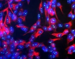
![Fibrillarin + FBLL1 antibody [HL2588] detects Fibrillarin + FBLL1 protein at nuclear envelope and nucleus by immunofluorescent analysis. Sample: HeLa cells were fixed in 4% paraformaldehyde at RT for 15 min. Green: Fibrillarin + FBLL1 stained by Fibrillarin + FBLL1 antibody [HL2588] (GTX639039) diluted at 1:500. Red: alpha Tubulin, a cytoskeleton marker, stained by alpha Tubulin antibody [GT114] (GTX628802) diluted at 1:1000. Blue: Fluoroshield with DAPI (GTX30920).](https://www.genetex.com/upload/website/prouct_img/normal/GTX639039/GTX639039_T-45159_20231110_ICC_IF_24051400_571.webp)
Product group Antibodies
ApplicationsImmunoFluorescence, Western Blot, ImmunoCytoChemistry, ImmunoHistoChemistry, ImmunoHistoChemistry Paraffin
ReactivityHuman, Mouse, Rat
TargetFBL
- SizePrice