

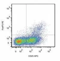
Intracellular staining of human peripheral blood cells (gated on CD4+ cells) with anti-FoxP3 (clone 3G3).
 with anti-FoxP3 (clone 3G3) (GTX80283)")
Intracellular staining of human peripheral blood cells (gated on CD4+ cells) with anti-FoxP3 (clone 3G3).
FOXP3 antibody [3G3]
GTX80283
ApplicationsFlow Cytometry, Western Blot
Product group Antibodies
ReactivityHuman, Mouse
TargetFoxp3
Overview
- SupplierGeneTex
- Product NameFOXP3 antibody [3G3]
- Delivery Days Customer9
- ApplicationsFlow Cytometry, Western Blot
- CertificationResearch Use Only
- ClonalityMonoclonal
- Clone ID3G3
- Concentration1 mg/ml
- ConjugateUnconjugated
- Gene ID20371
- Target nameFoxp3
- Target descriptionforkhead box P3
- Target synonymsJM2, scurfin, sf, forkhead box protein P3, scurfy
- HostMouse
- IsotypeIgG1
- Protein IDQ99JB6
- Protein NameForkhead box protein P3
- Scientific DescriptionThe protein encoded by this gene is a member of the forkhead/winged-helix family of transcriptional regulators. Defects in this gene result in the scurfy phenotype (sf). Alternative splicing results in multiple transcript variants. [provided by RefSeq, Sep 2015]
- ReactivityHuman, Mouse
- Storage Instruction2°C to 8°C
- UNSPSC41116161
Datasheet
Related products
Product group Antibodies
anti-FOXP3 (mouse), pAbAG-25A-0020
ApplicationsWestern Blot, ELISA
ReactivityMouse
TargetFoxp3
- SizePrice

Product group Antibodies
Anti-Foxp3 Antibody Picoband(r)A00011-2-CARRIER-FREE
ApplicationsWestern Blot
ReactivityMouse, Rat
TargetFoxp3
- SizePrice
Product group Antibodies

ApplicationsFlow Cytometry, ImmunoFluorescence, ELISA, ImmunoHistoChemistry
ReactivityMouse, Rat
TargetFoxp3
- SizePrice
Product group Antibodies
ApplicationsImmunoPrecipitation, Western Blot, ImmunoCytoChemistry, ImmunoHistoChemistry
ReactivityMouse
TargetFoxp3
- SizePrice
![FACS analysis of mouse C57Bl/6 splenocytes using GTX01488-07 FOXP3 antibody [MF23] (APC). Right panel : primary antibody Left panel : isotype control antibody amount : 0.25 μg (5 μl)](https://www.genetex.com/upload/website/prouct_img/normal/GTX01488-07/GTX01488-07_20200428_FACS9_w_23053121_490.webp)
Product group Antibodies
FOXP3 antibody [MF23] (APC)GTX01488-07
ApplicationsFlow Cytometry
ReactivityMouse
TargetFoxp3
- SizePrice
![FACS analysis of mouse C57Bl/6 splenocytes using GTX01488-08 FOXP3 antibody [MF23] (PE). Right panel : primary antibody Left panel : isotype control antibody amount : 0.25 μg (5 μl)](https://www.genetex.com/upload/website/prouct_img/normal/GTX01488-08/GTX01488-08_20200428_FACS99_w_23053121_811.webp)
Product group Antibodies
FOXP3 antibody [MF23] (PE)GTX01488-08
ApplicationsFlow Cytometry
ReactivityMouse
TargetFoxp3
- SizePrice
![FACS analysis of C57Bl/6 splenocytes stained FITC anti-mouse CD4 antibody, followed by intracellular staining with GTX80283-07 FOXP3 antibody [3G3] (APC). Right panel : Co-stained with FITC anti-mouse CD4 antibody and GTX80283-07 Left panel : Co-stained with FITC anti-mouse CD4 antibody and APC mouse IgG1 isotype control Antibody amount : 0.125 μg](https://www.genetex.com/upload/website/prouct_img/normal/GTX80283-07/GTX80283-07_20201020_FACS_11_w_23061322_251.webp)
Product group Antibodies
FOXP3 antibody [3G3] (APC)GTX80283-07
ApplicationsFlow Cytometry
ReactivityHuman, Mouse, Primate
TargetFoxp3
- SizePrice
![FACS analysis of C57Bl/6 splenocytes stained APC anti-mouse CD4 antibody, followed by intracellular staining with GTX80284 FOXP3 antibody [3G3] (PE). Right panel : Co-stained with APC anti-mouse CD4 antibody and GTX80284 Left panel : Co-stained with APC anti-mouse CD4 antibody and PE mouse IgG1 isotype control Antibody amount : 0.125 μg](https://www.genetex.com/upload/website/prouct_img/normal/GTX80284/GTX80284_20201020_FACS_64_w_23061322_985.webp)
Product group Antibodies
FOXP3 antibody [3G3] (PE)GTX80284
ApplicationsFlow Cytometry
ReactivityHuman, Mouse, Primate
TargetFoxp3
- SizePrice
Product group Antibodies
References
FOXP3 antibody, C-termGTX89752
ApplicationsImmunoHistoChemistry, ImmunoHistoChemistry Paraffin
ReactivityMouse
TargetFoxp3
- SizePrice
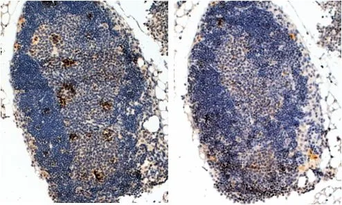
![FOXP3 antibody [C3], C-term detects FOXP3 protein at cytoplasm and nucleus in mouse spleen by immunohistochemical analysis. Sample: Paraffin-embedded mouse spleen. FOXP3 antibody [C3], C-term (GTX107737) diluted at 1:500.
Antigen Retrieval: Citrate buffer, pH 6.0, 15 min](https://www.genetex.com/upload/website/prouct_img/normal/GTX107737/GTX107737_39715_20170929_IHC-P_M_w_23060120_374.webp)
Product group Antibodies
FOXP3 antibody [C3], C-termGTX107737
ApplicationsImmunoFluorescence, Western Blot, ImmunoCytoChemistry, ImmunoHistoChemistry, ImmunoHistoChemistry Paraffin
ReactivityHuman, Mouse
TargetFoxp3
- SizePrice