

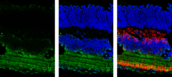
GAD65 antibody [C2C3], C-term detects GAD65 protein by immunohistochemical analysis. Sample: Frozen sectioned adult mouse retina. Green: GAD65 protein stained by GAD65 antibody [C2C3], C-term (GTX100281) diluted at 1:250. Red: Protein kinase C alpha staining. Blue: Fluoroshield with DAPI (GTX30920).
![GAD65 antibody [C2C3], C-term detects GAD2 protein at cytosol on RT2 xenograft by immunohistochemical analysis. Sample: Paraffin-embedded RT2 xenograft. GAD65 antibody [C2C3], C-term (GTX100281) dilution: 1:500.
Antigen Retrieval: Trilogy? (EDTA based, pH 8.0) buffer, 15min](https://www.genetex.com/upload/website/prouct_img/normal/GTX100281/GTX100281_39456_IHC_R_w_23060100_304.webp "GAD65 antibody [C2C3], C-term detects GAD2 protein at cytosol on RT2 xenograft by immunohistochemical analysis. Sample: Paraffin-embedded RT2 xenograft. GAD65 antibody [C2C3], C-term (GTX100281) dilution: 1:500.
Antigen Retrieval: Trilogy? (EDTA based, pH 8.0) buffer, 15min")
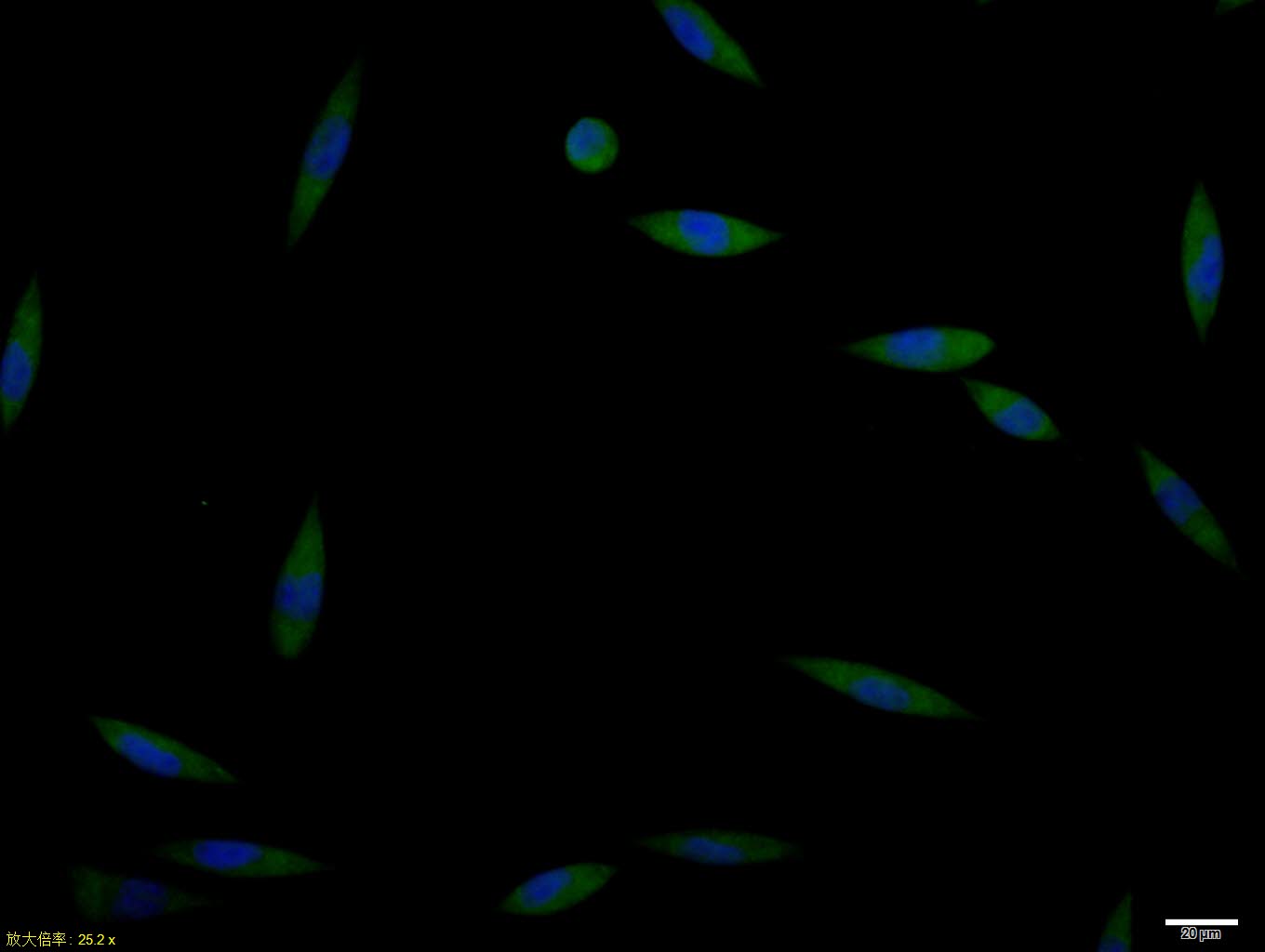
![GAD65 antibody [C2C3], C-term detects GAD65 protein expression by immunofluorescent analysis. Sample: Cultured Rat E18 primary cortical neuron, DIV 8. Cells were fixed in 4% paraformaldehyde at RT for 15 min. Red: GAD65 protein stained by GAD65 antibody [C2C3], C-term (GTX100281) diluted at 1:250. Blue: Fluoroshield with DAPI (GTX30920)..](https://www.genetex.com/upload/website/prouct_img/normal/GTX100281/GTX100281_39456_20161004_IFA_w_23060100_137.webp "GAD65 antibody [C2C3], C-term detects GAD65 protein expression by immunofluorescent analysis. Sample: Cultured Rat E18 primary cortical neuron, DIV 8. Cells were fixed in 4% paraformaldehyde at RT for 15 min. Red: GAD65 protein stained by GAD65 antibody [C2C3], C-term (GTX100281) diluted at 1:250. Blue: Fluoroshield with DAPI (GTX30920)..")
![Non-transfected (–) and transfected (+) 293T whole cell extracts (30 μg) were separated by 10% SDS-PAGE, and the membrane was blotted with GAD65 antibody [C2C3], C-term (GTX100281) diluted at 1:100000. The HRP-conjugated anti-rabbit IgG antibody (GTX213110-01) was used to detect the primary antibody.](https://www.genetex.com/upload/website/prouct_img/normal/GTX100281/GTX100281_39456_20220506_WB_B_w_23060100_348.webp "Non-transfected (–) and transfected (+) 293T whole cell extracts (30 μg) were separated by 10% SDS-PAGE, and the membrane was blotted with GAD65 antibody [C2C3], C-term (GTX100281) diluted at 1:100000. The HRP-conjugated anti-rabbit IgG antibody (GTX213110-01) was used to detect the primary antibody.")
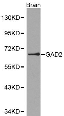
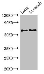
![Various tissue extracts (50 μg) were separated by 7.5% SDS-PAGE, and the membrane was blotted with GAD65 antibody [C2C3], C-term (GTX100281) diluted at 1:5000. The HRP-conjugated anti-rabbit IgG antibody (GTX213110-01) was used to detect the primary antibody.](https://www.genetex.com/upload/website/prouct_img/normal/GTX100281/GTX100281_39456_20180119_WB_M_R_w_23060100_455.webp "Various tissue extracts (50 μg) were separated by 7.5% SDS-PAGE, and the membrane was blotted with GAD65 antibody [C2C3], C-term (GTX100281) diluted at 1:5000. The HRP-conjugated anti-rabbit IgG antibody (GTX213110-01) was used to detect the primary antibody.")
GAD65 antibody [C2C3], C-term detects GAD65 protein by immunohistochemical analysis. Sample: Frozen sectioned adult mouse retina. Green: GAD65 protein stained by GAD65 antibody [C2C3], C-term (GTX100281) diluted at 1:250. Red: Protein kinase C alpha staining. Blue: Fluoroshield with DAPI (GTX30920).
GAD65 antibody [C2C3], C-term
GTX100281
ApplicationsImmunoFluorescence, Western Blot, ImmunoCytoChemistry, ImmunoHistoChemistry, ImmunoHistoChemistry Frozen, ImmunoHistoChemistry Paraffin
Product group Antibodies
ReactivityHuman, Mouse, Rat
TargetGAD2
Overview
- SupplierGeneTex
- Product NameGAD65 antibody [C2C3], C-term
- Delivery Days Customer9
- Application Supplier NoteWB: 1:1000-1:10000. ICC/IF: 1:100-1:1000. IHC-P: 1:100-1:1000. IHC-Fr: 1:100-1:1000. *Optimal dilutions/concentrations should be determined by the researcher.Not tested in other applications.
- ApplicationsImmunoFluorescence, Western Blot, ImmunoCytoChemistry, ImmunoHistoChemistry, ImmunoHistoChemistry Frozen, ImmunoHistoChemistry Paraffin
- CertificationResearch Use Only
- ClonalityPolyclonal
- Concentration1 mg/ml
- ConjugateUnconjugated
- Gene ID2572
- Target nameGAD2
- Target descriptionglutamate decarboxylase 2
- Target synonymsGAD65, glutamate decarboxylase 2, 65 kDa glutamic acid decarboxylase, GAD-65, Glutamate decarboxylase-2 (pancreas), glutamate decarboxylase 2 (pancreatic islets and brain, 65kDa)
- HostRabbit
- IsotypeIgG
- Protein IDQ05329
- Protein NameGlutamate decarboxylase 2
- Scientific DescriptionThis gene encodes one of several forms of glutamic acid decarboxylase, identified as a major autoantigen in insulin-dependent diabetes. The enzyme encoded is responsible for catalyzing the production of gamma-aminobutyric acid from L-glutamic acid. A pathogenic role for this enzyme has been identified in the human pancreas since it has been identified as an autoantibody and an autoreactive T cell target in insulin-dependent diabetes. This gene may also play a role in the stiff man syndrome. Alternative splicing results in multiple transcript variants that encode the same protein. [provided by RefSeq]
- ReactivityHuman, Mouse, Rat
- Storage Instruction-20°C or -80°C,2°C to 8°C
- UNSPSC41116161
Datasheet
Related products
Product group Antibodies
Anti-GAD2 AntibodyA28907
ApplicationsWestern Blot, ImmunoHistoChemistry
ReactivityHuman, Mouse, Rat
- SizePrice

Product group Antibodies
Anti-GAD65/GAD2 Antibody Picoband(r)A03142-1-CARRIER-FREE
ApplicationsWestern Blot, ELISA, ImmunoHistoChemistry
ReactivityHuman, Mouse, Rat
TargetGAD2
- SizePrice
Product group Antibodies
Anti-GAD2 Antibody144-00971
ApplicationsImmunoFluorescence, Western Blot, ImmunoHistoChemistry
ReactivityHuman, Mouse, Rat
TargetGAD2
- SizePrice

Product group Antibodies
Anti-GAD2 AntibodyAMAB91048
ApplicationsWestern Blot, ImmunoHistoChemistry
ReactivityHuman, Mouse, Rat
TargetGAD2
- SizePrice
Product group Antibodies
References
GAD65 Polyclonal AntibodyBS-0400R
ApplicationsFlow Cytometry, ImmunoFluorescence, Western Blot, ELISA, ImmunoCytoChemistry, ImmunoHistoChemistry, ImmunoHistoChemistry Frozen, ImmunoHistoChemistry Paraffin
ReactivityBovine, Canine, Chicken, Human, Mouse, Porcine, Rat
TargetGAD2
- SizePrice
Product group Antibodies
GAD2 AntibodyCSB-PA11159A0RB
ApplicationsWestern Blot, ELISA, ImmunoHistoChemistry
ReactivityHuman, Mouse
TargetGAD2
- SizePrice
Product group Antibodies

Goat anti-GAD2 / GAD65EB06730
ApplicationsWestern Blot, ELISA, ImmunoHistoChemistry
ReactivityCanine, Human, Mouse, Rat
TargetGAD2
- SizePrice
Product group Antibodies
Gad2 Polyclonal AntibodyCAC08038
ApplicationsWestern Blot, ELISA, ImmunoHistoChemistry
ReactivityMouse
TargetGAD2
- SizePrice
Product group Antibodies

GAD65 AntibodyLS-C405593
ApplicationsELISA, ImmunoHistoChemistry
ReactivityHuman
TargetGAD2
- SizePrice
Product group Antibodies
GAD65 antibody [GAD2/1960]GTX17966
ApplicationsImmunoHistoChemistry, ImmunoHistoChemistry Paraffin, Other Application
ReactivityHuman
TargetGAD2
- SizePrice