

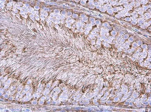
GAPDH antibody [GT239] (HRP) detects GAPDH antibody protein at cytoplasm in rat testis by immunohistochemical analysis. Sample: Paraffin-embedded rat testis. GAPDH antibody [GT239] (HRP) (GTX627408-01) diluted at 1:200.
Antigen Retrieval: Citrate buffer, pH 6.0, 15 min
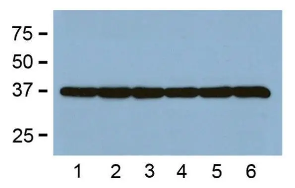
![Various whole cell extracts (30 μg) were separated by 10% SDS-PAGE, and the membrane was blotted with GAPDH antibody [GT239] (HRP) (GTX627408-01) diluted at 1:10000. The HRP-conjugated anti-rabbit IgG antibody (GTX213110-01) was used to detect the primary antibody.](https://www.genetex.com/upload/website/prouct_img/normal/GTX627408-01/GTX627408-01_44405_20210806_WB_w_23061202_657.webp "Various whole cell extracts (30 μg) were separated by 10% SDS-PAGE, and the membrane was blotted with GAPDH antibody [GT239] (HRP) (GTX627408-01) diluted at 1:10000. The HRP-conjugated anti-rabbit IgG antibody (GTX213110-01) was used to detect the primary antibody.")
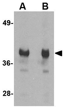
![Whole cell extract (30 μg) was separated by 10% SDS-PAGE, and the membrane was blotted with GAPDH antibody [GT239] (GTX627408) diluted at 1:1000. The HRP-conjugated anti-rabbit IgG antibody (GTX213110-01) was used to detect the primary antibody.](https://www.genetex.com/upload/website/prouct_img/normal/GTX627408-01/GTX627408_44405_20240126_WB_Hamster_24061301_264.webp "Whole cell extract (30 μg) was separated by 10% SDS-PAGE, and the membrane was blotted with GAPDH antibody [GT239] (GTX627408) diluted at 1:1000. The HRP-conjugated anti-rabbit IgG antibody (GTX213110-01) was used to detect the primary antibody.")
![Whole cell extract (30 μg) was separated by 10% SDS-PAGE, and the membrane was blotted with GAPDH antibody [GT239] (GTX627408) diluted at 1:1000. The HRP-conjugated anti-rabbit IgG antibody (GTX213110-01) was used to detect the primary antibody.](https://www.genetex.com/upload/website/prouct_img/normal/GTX627408-01/GTX627408_44405_20240126_WB_Hamster_2_24061301_301.webp "Whole cell extract (30 μg) was separated by 10% SDS-PAGE, and the membrane was blotted with GAPDH antibody [GT239] (GTX627408) diluted at 1:1000. The HRP-conjugated anti-rabbit IgG antibody (GTX213110-01) was used to detect the primary antibody.")
GAPDH antibody [GT239] (HRP) detects GAPDH antibody protein at cytoplasm in rat testis by immunohistochemical analysis. Sample: Paraffin-embedded rat testis. GAPDH antibody [GT239] (HRP) (GTX627408-01) diluted at 1:200.
Antigen Retrieval: Citrate buffer, pH 6.0, 15 min
GAPDH antibody [GT239] (HRP)
GTX627408-01
ApplicationsWestern Blot, ImmunoHistoChemistry, ImmunoHistoChemistry Paraffin
Product group Antibodies
ReactivityHamster, Human, Mouse, Rat
TargetGAPDH
Overview
- SupplierGeneTex
- Product NameGAPDH antibody [GT239] (HRP)
- Delivery Days Customer9
- Application Supplier NoteWB: 1:5000-1:20000. IHC-P: 1:100-1:1000. *Optimal dilutions/concentrations should be determined by the researcher.Not tested in other applications.
- ApplicationsWestern Blot, ImmunoHistoChemistry, ImmunoHistoChemistry Paraffin
- CertificationResearch Use Only
- ClonalityMonoclonal
- Clone IDGT239
- Concentration0.62 mg/ml
- ConjugateHRP
- Gene ID2597
- Target nameGAPDH
- Target descriptionglyceraldehyde-3-phosphate dehydrogenase
- Target synonymsG3PD, GAPD, HEL-S-162eP, glyceraldehyde-3-phosphate dehydrogenase, OCAS, p38 component, Oct1 coactivator in S phase, 38 Kd component, aging-associated gene 9 protein, epididymis secretory sperm binding protein Li 162eP, peptidyl-cysteine S-nitrosylase GAPDH
- HostMouse
- IsotypeIgG2b
- Protein IDP04406
- Protein NameGlyceraldehyde-3-phosphate dehydrogenase
- Scientific DescriptionThe product of this gene catalyzes an important energy-yielding step in carbohydrate metabolism, the reversible oxidative phosphorylation of glyceraldehyde-3-phosphate in the presence of inorganic phosphate and nicotinamide adenine dinucleotide (NAD). The enzyme exists as a tetramer of identical chains. Many pseudogenes similar to this locus are present in the human genome. [provided by RefSeq]
- ReactivityHamster, Human, Mouse, Rat
- Storage Instruction-20°C or -80°C,2°C to 8°C
- UNSPSC12352203
References
- Zengel J, Wang YX, Seo JW, et al. Hardwiring tissue-specific AAV transduction in mice through engineered receptor expression. Nat Methods. 2023,20(7):1070-1081. doi: 10.1038/s41592-023-01896-xRead this paper
- Escalante-Covarrubias Q, Mendoza-Viveros L, González-Suárez M, et al. Time-of-day defines NAD(+) efficacy to treat diet-induced metabolic disease by synchronizing the hepatic clock in mice. Nat Commun. 2023,14(1):1685. doi: 10.1038/s41467-023-37286-2Read this paper
- Ferdous S, Shelton DA, Getz TE, et al. Deletion of histone demethylase Lsd1 (Kdm1a) during retinal development leads to defects in retinal function and structure. Front Cell Neurosci. 2023,17:1104592. doi: 10.3389/fncel.2023.1104592Read this paper
- Cheng H, Zhang Y, Guo X, et al. Study on Neuroprotective Mechanism of Houshiheisan in Ischemic Stroke Based on Transcriptomics and Experimental Verification. Evid Based Complement Alternat Med. 2023,2023:8673136. doi: 10.1155/2023/8673136Read this paper
- Shilts J, Crozier TWM, Teixeira-Silva A, et al. LRRC15 mediates an accessory interaction with the SARS-CoV-2 spike protein. PLoS Biol. 2023,21(2):e3001959. doi: 10.1371/journal.pbio.3001959Read this paper
- Despic V, Jaffrey SR. mRNA ageing shapes the Cap2 methylome in mammalian mRNA. Nature. 2023,614(7947):358-366. doi: 10.1038/s41586-022-05668-zRead this paper
- Quiles JM, Najor RH, Gonzalez E, et al. Deciphering functional roles and interplay between Beclin1 and Beclin2 in autophagosome formation and mitophagy. Sci Signal. 2023,16(770):eabo4457. doi: 10.1126/scisignal.abo4457Read this paper
- Chien HM, He RY, Lee CC, et al. Nanoscopic investigation of C9orf72 poly-GA oligomers on nuclear membrane disruption by a photoinducible platform. Commun Chem. 2021,4(1):111. doi: 10.1038/s42004-021-00547-6Read this paper
- Arimoto KI, Miyauchi S, Troutman TD, et al. Expansion of interferon inducible gene pool via USP18 inhibition promotes cancer cell pyroptosis. Nat Commun. 2023,14(1):251. doi: 10.1038/s41467-022-35348-5Read this paper
- Sych K, Nold SP, Pfeilschifter J, et al. Expression of PKM2 in wound keratinocytes is coupled to angiogenesis during skin repair in vivo and in HaCaT keratinocytes in vitro. J Mol Med (Berl). 2023,101(1-2):151-169. doi: 10.1007/s00109-022-02280-6Read this paper
Datasheet
Related products
Product group Antibodies
Anti-GAPDH (63~83aa) Antibody130-10873
ApplicationsELISA
ReactivityHuman, Rabbit
TargetGAPDH
- SizePrice


Product group Antibodies
Anti-GAPDH Antibody Picoband(r)A00227-1-CARRIER-FREE
ApplicationsFlow Cytometry, ImmunoFluorescence, Western Blot, ImmunoCytoChemistry, ImmunoHistoChemistry
ReactivityChicken, Human, Monkey, Mouse, Rat, Zebra Fish
TargetGAPDH
- SizePrice
![ICC/IF analysis of NIH-3T3 cells using GTX03407 GAPDH antibody [GT1355]. Red : Primary antibody Blue : DAPI for nuclear staining Dilution : 1:100](https://www.genetex.com/upload/website/prouct_img/normal/GTX03407/GTX03407_ICCIF_1_w_23053123_726.webp)
Product group Antibodies
GAPDH antibody [GT1355]GTX03407
ApplicationsImmunoFluorescence, Western Blot, ImmunoCytoChemistry, ImmunoHistoChemistry, ImmunoHistoChemistry Paraffin
ReactivityHuman, Mouse, Rat
TargetGAPDH
- SizePrice
![Various whole cell extracts (30 μg) were separated by 10% SDS-PAGE, and the membrane was blotted with GAPDH antibody [HL2062] (GTX637966) diluted at 1:5000. The HRP-conjugated anti-rabbit IgG antibody (GTX213110-01) was used to detect the primary antibody.](https://www.genetex.com/upload/website/prouct_img/normal/GTX637966/GTX637966_T-44886_20221209_WB_M_R_23010400_421.webp)
Product group Antibodies
GAPDH antibody [HL2062]GTX637966
ApplicationsImmunoFluorescence, Western Blot, ImmunoCytoChemistry
ReactivityBacteria, Canine, Feline, Hamster, Human, Monkey, Mouse, Rabbit, Rat, Zebra Fish
TargetGAPDH
- SizePrice
Product group Antibodies
References
GAPDH antibody [GA1R]GTX82560
ApplicationsImmunoFluorescence, Western Blot, ELISA, ImmunoCytoChemistry, ImmunoHistoChemistry
ReactivityBacteria, Chicken, Hamster, Human, Mouse, Rabbit, Rat, Yeast
TargetGAPDH
- SizePrice
![WB analysis of HeLa (1), A549 (2), A431 (3), MCF-7 (4), K562 (5), Jurkat (6), HL60 (7), SKN-SH (8) and SKBR-3 (9) cell lysate using GTX83245 GAPDH antibody [1A10].](https://www.genetex.com/upload/website/prouct_img/normal/GTX83245/GTX83245_20170912_WB_w_23061322_915.webp)
Product group Antibodies
GAPDH antibody [1A10]GTX83245
ApplicationsImmunoFluorescence, Western Blot, ELISA, ImmunoCytoChemistry, ImmunoHistoChemistry, ImmunoHistoChemistry Paraffin
ReactivityHuman
TargetGAPDH
- SizePrice
Product group Antibodies
References
GAPDH antibodyGTX85118
ApplicationsWestern Blot, ELISA
ReactivityHuman, Mouse, Rat
TargetGAPDH
- SizePrice
Product group Antibodies
Gapdh Polyclonal AntibodyCAC07001
ApplicationsImmunoFluorescence, Western Blot, ELISA, ImmunoHistoChemistry
TargetGAPDH
- SizePrice