

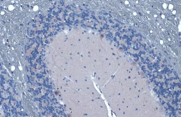
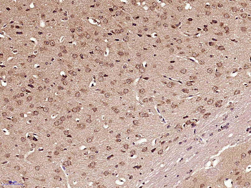
Gli1 antibody detects Gli1 protein at cytoplasm and nucleus by immunohistochemical analysis. Sample: Paraffin-embedded mouse cerebellum. Gli1 stained by Gli1 antibody (GTX106207) diluted at 1:500. Antigen Retrieval: Citrate buffer, pH 6.0, 15 min
 was separated by 5% SDS-PAGE, and the membrane was blotted with Gli1 antibody (GTX106207) diluted at 1:1000. The HRP-conjugated anti-rabbit IgG antibody (GTX213110-01) was used to detect the primary antibody.")
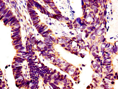
 diluted at 1:500.")
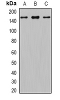
 were separated by 5% SDS-PAGE, and the membrane was blotted with Gli1 antibody (GTX106207) diluted at 1:500. The HRP-conjugated anti-rabbit IgG antibody (GTX213110-01) was used to detect the primary antibody.")
. Western blot analysis was performed using Gli1 antibody (GTX106207) diluted at 1:500. EasyBlot HRP-conjugated anti rabbit IgG antibody (GTX221666-01) was used to detect the primary antibody.")
 and transfected (+) 293T whole cell extracts (30 μg) were separated by 5% SDS-PAGE, and the membrane was blotted with Gli1 antibody (GTX106207) diluted at 1:5000. The HRP-conjugated anti-rabbit IgG antibody (GTX213110-01) was used to detect the primary antibody.")
Gli1 antibody detects Gli1 protein at cytoplasm and nucleus by immunohistochemical analysis. Sample: Paraffin-embedded mouse cerebellum. Gli1 stained by Gli1 antibody (GTX106207) diluted at 1:500. Antigen Retrieval: Citrate buffer, pH 6.0, 15 min
Gli1 antibody
GTX106207
ApplicationsImmunoFluorescence, ImmunoPrecipitation, Western Blot, ImmunoCytoChemistry, ImmunoHistoChemistry, ImmunoHistoChemistry Paraffin
Product group Antibodies
ReactivityHuman, Mouse
TargetGLI1
Overview
- SupplierGeneTex
- Product NameGli1 antibody
- Delivery Days Customer9
- Application Supplier NoteWB: 1:500-1:10000. ICC/IF: 1:100-1:1000. IP: 1:1000-1:1000. *Optimal dilutions/concentrations should be determined by the researcher.Not tested in other applications.
- ApplicationsImmunoFluorescence, ImmunoPrecipitation, Western Blot, ImmunoCytoChemistry, ImmunoHistoChemistry, ImmunoHistoChemistry Paraffin
- CertificationResearch Use Only
- ClonalityPolyclonal
- Concentration1.12 mg/ml
- ConjugateUnconjugated
- Gene ID2735
- Target nameGLI1
- Target descriptionGLI family zinc finger 1
- Target synonymsGLI, PAPA8, PPD1, zinc finger protein GLI1, GLI-Kruppel family member GLI1, glioma-associated oncogene 1, glioma-associated oncogene homolog 1 (zinc finger protein), oncogene GLI
- HostRabbit
- IsotypeIgG
- Protein IDP08151
- Protein NameZinc finger protein GLI1
- Scientific DescriptionThis gene encodes a protein which is a member of the Kruppel family of zinc finger proteins. The function of this gene has not been determined; however, it may play a role in normal development gene transcription. Mouse mutation studies indicate possible involvement in human foregut malformation. [provided by RefSeq]
- ReactivityHuman, Mouse
- Storage Instruction-20°C or -80°C,2°C to 8°C
- UNSPSC41116161
Datasheet
Related products
Product group Antibodies
Anti-Gli1 AntibodyA121161
ApplicationsFlow Cytometry, ImmunoFluorescence, ELISA
ReactivityHuman
- SizePrice
Product group Antibodies
Anti-GLI1 Antibody144-08387
ApplicationsWestern Blot
ReactivityHuman, Mouse, Rat
TargetGLI1
- SizePrice

Product group Antibodies
Anti-GLI1 AntibodyAMAB91771
ApplicationsWestern Blot, ImmunoCytoChemistry
ReactivityHuman
TargetGLI1
- SizePrice
Product group Antibodies

GLI / GLI1 AntibodyLS-C834930
ApplicationsWestern Blot, ELISA, ImmunoHistoChemistry
ReactivityHuman, Mouse
TargetGLI1
- SizePrice

Product group Antibodies
Anti-GLI1 Antibody Picoband(r)A00527-3-CARRIER-FREE
ApplicationsWestern Blot, ELISA
ReactivityHuman, Mouse, Rat
TargetGLI1
- SizePrice
Product group Antibodies
References
GLI1 Polyclonal AntibodyBS-1206R
ApplicationsFlow Cytometry, ImmunoFluorescence, Western Blot, ELISA, ImmunoCytoChemistry, ImmunoHistoChemistry, ImmunoHistoChemistry Frozen, ImmunoHistoChemistry Paraffin
ReactivityBovine, Canine, Equine, Human, Mouse, Rabbit, Rat
TargetGLI1
- SizePrice
Product group Antibodies
GLI1 AntibodyCSB-PA12989A0RB
ApplicationsImmunoFluorescence, ELISA, ImmunoHistoChemistry
ReactivityHuman
TargetGLI1
- SizePrice
Product group Antibodies

Goat anti-GLI1EB10594
ApplicationsFlow Cytometry, ImmunoFluorescence, ELISA
ReactivityBovine, Canine, Human, Mouse, Rat
TargetGLI1
- SizePrice
Product group Antibodies
References
Gli1 antibodyGTX17495
ApplicationsWestern Blot
ReactivityHuman, Mouse, Rat
TargetGLI1
- SizePrice
![Whole cell extract (30 μg) was separated by 5% SDS-PAGE, and the membrane was blotted with Gli1 antibody [GT249] (GTX635705) diluted at 1:1000. The HRP-conjugated anti-mouse IgG antibody (GTX213111-01) was used to detect the primary antibody, and the signal was developed with Trident ECL plus-Enhanced.](https://www.genetex.com/upload/website/prouct_img/normal/GTX635705/GTX635705_44004_20200717_WB_w_23061202_283.webp)
Product group Antibodies
Gli1 antibody [GT249]GTX635705
ApplicationsWestern Blot, ImmunoHistoChemistry, ImmunoHistoChemistry Paraffin
ReactivityHuman, Mouse
TargetGLI1
- SizePrice