

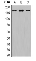
WB analysis of PC3 (A), mouse brain (B), rat brain (C) whole cell lysates using GTX17495 Gli1 antibody.
WB analysis of PC3 (A), mouse brain (B), rat brain (C) whole cell lysates using GTX17495 Gli1 antibody.
Gli1 antibody
GTX17495
ApplicationsWestern Blot
Product group Antibodies
ReactivityHuman, Mouse, Rat
TargetGLI1
Overview
- SupplierGeneTex
- Product NameGli1 antibody
- Delivery Days Customer9
- Application Supplier NoteWB: 1:500 - 1:2000. *Optimal dilutions/concentrations should be determined by the researcher.Not tested in other applications.
- ApplicationsWestern Blot
- CertificationResearch Use Only
- ClonalityPolyclonal
- ConjugateUnconjugated
- Gene ID2735
- Target nameGLI1
- Target descriptionGLI family zinc finger 1
- Target synonymsGLI, PAPA8, PPD1, zinc finger protein GLI1, GLI-Kruppel family member GLI1, glioma-associated oncogene 1, glioma-associated oncogene homolog 1 (zinc finger protein), oncogene GLI
- HostRabbit
- IsotypeIgG
- Protein IDP08151
- Protein NameZinc finger protein GLI1
- Scientific DescriptionThis gene encodes a member of the Kruppel family of zinc finger proteins. The encoded transcription factor is activated by the sonic hedgehog signal transduction cascade and regulates stem cell proliferation. The activity and nuclear localization of this protein is negatively regulated by p53 in an inhibitory loop. Multiple transcript variants encoding different isoforms have been found for this gene. [provided by RefSeq, May 2009]
- ReactivityHuman, Mouse, Rat
- Storage Instruction-20°C or -80°C,2°C to 8°C
- UNSPSC41116161
References
- ITR-284 modulates cell differentiation in human chronic myelogenous leukemia K562 cells. Yang JS et al., 2018 Jan, Oncol RepRead this paper
Datasheet
Related products
Product group Antibodies
Anti-Gli1 AntibodyA121161
ApplicationsFlow Cytometry, ImmunoFluorescence, ELISA
ReactivityHuman
- SizePrice
Product group Antibodies
Anti-GLI1 Antibody144-08387
ApplicationsWestern Blot
ReactivityHuman, Mouse, Rat
TargetGLI1
- SizePrice

Product group Antibodies
Anti-GLI1 AntibodyAMAB91771
ApplicationsWestern Blot, ImmunoCytoChemistry
ReactivityHuman
TargetGLI1
- SizePrice
Product group Antibodies

GLI / GLI1 AntibodyLS-C834930
ApplicationsWestern Blot, ELISA, ImmunoHistoChemistry
ReactivityHuman, Mouse
TargetGLI1
- SizePrice

Product group Antibodies
Anti-GLI1 Antibody Picoband(r)A00527-3-CARRIER-FREE
ApplicationsWestern Blot, ELISA
ReactivityHuman, Mouse, Rat
TargetGLI1
- SizePrice
Product group Antibodies
References
GLI1 Polyclonal AntibodyBS-1206R
ApplicationsFlow Cytometry, ImmunoFluorescence, Western Blot, ELISA, ImmunoCytoChemistry, ImmunoHistoChemistry, ImmunoHistoChemistry Frozen, ImmunoHistoChemistry Paraffin
ReactivityBovine, Canine, Equine, Human, Mouse, Rabbit, Rat
TargetGLI1
- SizePrice
Product group Antibodies
GLI1 AntibodyCSB-PA12989A0RB
ApplicationsImmunoFluorescence, ELISA, ImmunoHistoChemistry
ReactivityHuman
TargetGLI1
- SizePrice
Product group Antibodies

Goat anti-GLI1EB10594
ApplicationsFlow Cytometry, ImmunoFluorescence, ELISA
ReactivityBovine, Canine, Human, Mouse, Rat
TargetGLI1
- SizePrice
Product group Antibodies
Gli1 antibodyGTX106207
ApplicationsImmunoFluorescence, ImmunoPrecipitation, Western Blot, ImmunoCytoChemistry, ImmunoHistoChemistry, ImmunoHistoChemistry Paraffin
ReactivityHuman, Mouse
TargetGLI1
- SizePrice
![Whole cell extract (30 μg) was separated by 5% SDS-PAGE, and the membrane was blotted with Gli1 antibody [GT249] (GTX635705) diluted at 1:1000. The HRP-conjugated anti-mouse IgG antibody (GTX213111-01) was used to detect the primary antibody, and the signal was developed with Trident ECL plus-Enhanced.](https://www.genetex.com/upload/website/prouct_img/normal/GTX635705/GTX635705_44004_20200717_WB_w_23061202_283.webp)
Product group Antibodies
Gli1 antibody [GT249]GTX635705
ApplicationsWestern Blot, ImmunoHistoChemistry, ImmunoHistoChemistry Paraffin
ReactivityHuman, Mouse
TargetGLI1
- SizePrice