

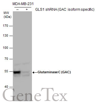
Glutaminase C (GAC) antibody validation by shRNA knock-down. Non-transfected (-) and GLS1(GAC isoform specific) shRNA-transfected MDA-MB-231 whole cell extracts (30 μg) were separated by 7.5% SDS-PAGE, and the membrane was blotted with Glutaminase C (GAC) antibody (GTX131263) at a dilution of 1:5000.
 or Glutaminase C antibody (GTX132402). Western blot analysis was performed using Glutaminase C antibody (GTX131263). EasyBlot anti-Rabbit IgG (GTX221666-01) was used as a secondary reagent.")
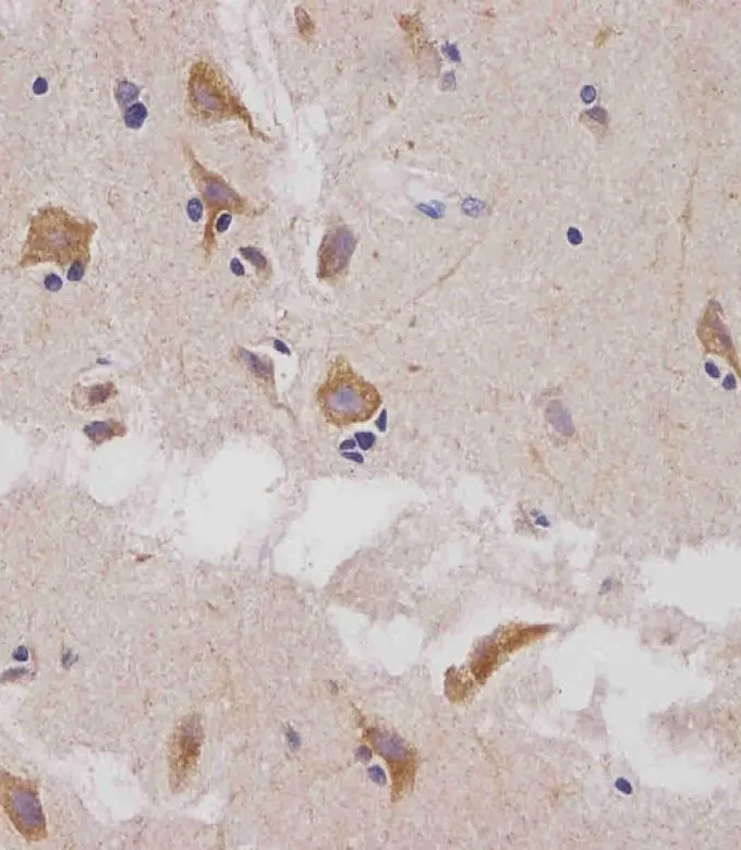
 diluted at 1:500.
Antigen Retrieval: Citrate buffer, pH 6.0, 15 min")
 diluted at 1:500.
Antigen Retrieval: Trilogy? (EDTA based, pH 8.0) buffer, 15min")
 diluted at 1:500.
Antigen Retrieval: Citrate buffer, pH 6.0, 15 min")
![Glutaminase C antibody detects Glutaminase C protein at mitochondria by immunofluorescent analysis. Sample: A549 cells were fixed in 4% paraformaldehyde at RT for 15 min. Green: Glutaminase C protein stained by Glutaminase C antibody (GTX131263) diluted at 1:500. Red: Histone H3K9ac (acetyl Lys9), a nucleus marker, stained by Histone H3K9ac (acetyl Lys9) antibody [GT464] (GTX630554) diluted at 1:500. Blue: Hoechst 33342 staining. Scale bar = 10 μm.](https://www.genetex.com/upload/website/prouct_img/normal/GTX131263/GTX131263_41976_20150410_IFA_w_23060523_623.webp "Glutaminase C antibody detects Glutaminase C protein at mitochondria by immunofluorescent analysis. Sample: A549 cells were fixed in 4% paraformaldehyde at RT for 15 min. Green: Glutaminase C protein stained by Glutaminase C antibody (GTX131263) diluted at 1:500. Red: Histone H3K9ac (acetyl Lys9), a nucleus marker, stained by Histone H3K9ac (acetyl Lys9) antibody [GT464] (GTX630554) diluted at 1:500. Blue: Hoechst 33342 staining. Scale bar = 10 μm.")
Glutaminase C (GAC) antibody validation by shRNA knock-down. Non-transfected (-) and GLS1(GAC isoform specific) shRNA-transfected MDA-MB-231 whole cell extracts (30 μg) were separated by 7.5% SDS-PAGE, and the membrane was blotted with Glutaminase C (GAC) antibody (GTX131263) at a dilution of 1:5000.
Glutaminase C (GAC) antibody
GTX131263
ApplicationsImmunoFluorescence, ImmunoPrecipitation, Western Blot, ImmunoCytoChemistry, ImmunoHistoChemistry, ImmunoHistoChemistry Paraffin
Product group Antibodies
ReactivityHuman, Mouse
TargetGLS
Overview
- SupplierGeneTex
- Product NameGlutaminase C (GAC) antibody
- Delivery Days Customer9
- Application Supplier NoteWB: 1:1000-1:10000. ICC/IF: 1:100-1:1000. IHC-P: 1:100-1:1000. IP: 1:100-1:500. *Optimal dilutions/concentrations should be determined by the researcher.Not tested in other applications.
- ApplicationsImmunoFluorescence, ImmunoPrecipitation, Western Blot, ImmunoCytoChemistry, ImmunoHistoChemistry, ImmunoHistoChemistry Paraffin
- CertificationResearch Use Only
- ClonalityPolyclonal
- Concentration0.5 mg/ml
- ConjugateUnconjugated
- Gene ID2744
- Target nameGLS
- Target descriptionglutaminase
- Target synonymsAAD20, CASGID, DEE71, EIEE71, GAC, GAM, GDPAG, GLS1, KGA, glutaminase kidney isoform, mitochondrial, K-glutaminase, L-glutamine amidohydrolase, glutaminase C, glutaminase, phosphate-activated
- HostRabbit
- IsotypeIgG
- Protein IDO94925
- Protein NameGlutaminase kidney isoform, mitochondrial
- Scientific DescriptionSahai (1983) [PubMed 6825316] demonstrated phosphate-activated glutaminase (EC 3.5.1.2) in human platelets. It is the major enzyme yielding glutamate from glutamine. Significance of the enzyme derives from its possible implication in behavior disturbances in which glutamate acts as a neurotransmitter (Prusiner, 1981). High heritability of platelet glutaminase was indicated by studies of Sahai and Vogel (1983) [PubMed 6682827] who found an intraclass correlation coefficient of 0.96 for monozygotic twins and 0.53 for dizygotic twins.[supplied by OMIM]
- ReactivityHuman, Mouse
- Storage Instruction-20°C or -80°C,2°C to 8°C
- UNSPSC12352203
References
- Barreca F, Aventaggiato M, Vitiello L, et al. SIRT5 Activation and Inorganic Phosphate Binding Reduce Cancer Cell Vitality by Modulating Autophagy/Mitophagy and ROS. Antioxidants (Basel). 2023,12(8). doi: 10.3390/antiox12081635Read this paper
- Fazal S, Danzi MC, van Kuilenburg ABP, et al. Repeat expansions nested within tandem CNVs: a unique structural change in GLS exemplifies the diagnostic challenges of non-coding pathogenic variation. Hum Mol Genet. 2023,32(1):46-54. doi: 10.1093/hmg/ddac173Read this paper
- Saladini S, Aventaggiato M, Barreca F, et al. Metformin Impairs Glutamine Metabolism and Autophagy in Tumour Cells. Cells. 2019,8(1). doi: 10.3390/cells8010049Read this paper
Datasheet
Related products
Product group Antibodies
Anti-GLS Antibody144-03885
ApplicationsWestern Blot, ImmunoHistoChemistry
ReactivityHuman, Mouse, Rat
TargetGLS
- SizePrice


Product group Antibodies
Anti-Glutaminase/GLS Antibody Picoband(r)A01272-2-CARRIER-FREE
ApplicationsFlow Cytometry, ImmunoFluorescence, Western Blot, ELISA, ImmunoCytoChemistry, ImmunoHistoChemistry
ReactivityHuman, Monkey, Mouse, Rat
TargetGLS
- SizePrice
![Glutaminase C antibody detects Glutaminase C protein at mitochondria by immunofluorescent analysis. Sample: A549 cells were fixed in 4% paraformaldehyde at RT for 15 min. Green: Glutaminase C protein stained by Glutaminase C antibody (GTX132402) diluted at 1:500. Red: Histone H3K9ac (acetyl Lys9), a nucleus marker, stained by Histone H3K9ac (acetyl Lys9) antibody [GT464] (GTX630554) diluted at 1:500. Blue: Hoechst 33342 staining. Scale bar = 10 μm.](https://www.genetex.com/upload/website/prouct_img/normal/GTX132402/GTX132402_41829_20150410_IFA_w_23060523_714.webp)
Product group Antibodies
Glutaminase C (GAC) antibodyGTX132402
ApplicationsImmunoFluorescence, ImmunoPrecipitation, Western Blot, ImmunoCytoChemistry, ImmunoHistoChemistry, ImmunoHistoChemistry Paraffin
ReactivityHuman
TargetGLS
- SizePrice
![Glutaminase antibody [C1C3] detects Glutaminase protein by Western blot analysis. A. 20 μg Raw264 whole cell lysate/extract Glutaminase antibody [C1C3] (GTX114982) dilution: 1:1000](https://www.genetex.com/upload/website/prouct_img/normal/GTX114982/GTX114982_40240_CT_WB_M_w_23060519_282.webp)
Product group Antibodies
Glutaminase antibody [C1C3]GTX114982
ApplicationsWestern Blot
ReactivityHuman, Mouse
TargetGLS
- SizePrice
![Glutaminase C antibody [GT1075] detects Glutaminase C protein at mitochondria by immunofluorescent analysis. Sample: HeLa cells were fixed in 4% paraformaldehyde at RT for 15 min. Green: Glutaminase C protein stained by Glutaminase C antibody [GT1075] (GTX632265) diluted at 1:400. Blue: Hoechst 33342 staining.](https://www.genetex.com/upload/website/prouct_img/normal/GTX632265/GTX632265_42030_20150831_IFA_w_23061202_626.webp)
Product group Antibodies
ApplicationsImmunoFluorescence, Western Blot, ImmunoCytoChemistry
ReactivityHuman
TargetGLS
- SizePrice
![Glutaminase C (GAC) antibody [GT3211] detects Glutaminase C (GAC) protein by western blot analysis. Various whole cell extracts (30 μg) were separated by 7.5% SDS-PAGE, and the membrane was blotted with Glutaminase C (GAC) antibody [GT3211] (GTX632317) diluted at a dilution of 1:1000.](https://www.genetex.com/upload/website/prouct_img/normal/GTX632317/GTX632317_42086_20150508_WB_w_23061202_695.webp)
Product group Antibodies
ApplicationsImmunoFluorescence, Western Blot, ImmunoCytoChemistry, ImmunoHistoChemistry, ImmunoHistoChemistry Paraffin
ReactivityHuman, Mouse
TargetGLS
- SizePrice
Product group Antibodies
References
Glutaminase antibody, C-termGTX81012
ApplicationsFlow Cytometry, ImmunoFluorescence, Western Blot, ImmunoCytoChemistry, ImmunoHistoChemistry, ImmunoHistoChemistry Paraffin
ReactivityHuman, Mouse, Rat
TargetGLS
- SizePrice
Product group Antibodies
GLS Polyclonal AntibodyCAC13047
ApplicationsWestern Blot, ELISA, ImmunoHistoChemistry
ReactivityMouse
TargetGLS
- SizePrice