

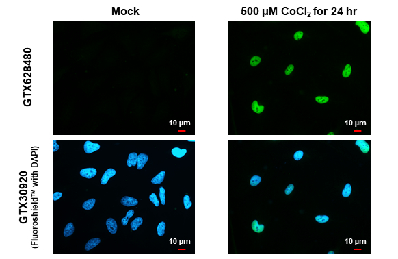
HIF1 alpha antibody [GT10211] detects HIF1 alpha protein at nucleus by immunofluorescent analysis. Sample: Mock and treated HeLa cells were fixed in 4% paraformaldehyde at RT for 15 min. Green: HIF1 alpha stained by HIF1 alpha antibody [GT10211] (GTX628480) diluted at 1:500. Blue: Fluoroshield with DAPI (GTX30920).
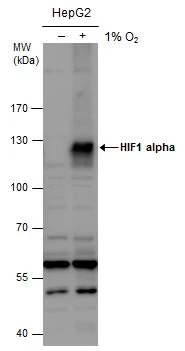
![HIF1 alpha antibody [GT10211] detects HIF1A protein by western blot analysis. A. 30 μg HepG2 whole cell lysate/extract (untreated) B. 30 μg HepG2 whole cell lysate/extract (1%O2 treatment for 48hr) 7.5% SDS-PAGE HIF1 alpha antibody [GT10211] (GTX628480) dilution: 1:250 The HRP-conjugated anti-mouse IgG antibody (GTX213111-01) was used to detect the primary antibody.](https://www.genetex.com/upload/website/prouct_img/normal/GTX628480/GTX628480_41134_WB_Hypoxia_w_23061202_732.webp "HIF1 alpha antibody [GT10211] detects HIF1A protein by western blot analysis. A. 30 μg HepG2 whole cell lysate/extract (untreated) B. 30 μg HepG2 whole cell lysate/extract (1%O2 treatment for 48hr) 7.5% SDS-PAGE HIF1 alpha antibody [GT10211] (GTX628480) dilution: 1:250 The HRP-conjugated anti-mouse IgG antibody (GTX213111-01) was used to detect the primary antibody.")
 C. Control with 2.5 μg of preimmune mouse IgG/HepG2 whole cell lysate/extract from normoxia D. Control with 2.5 μg of preimmune mouse IgG/ HepG2 whole cell lysate/extract from hypoxia 24hr (1%O2) E. Immunoprecipitation of HIF-1 alpha protein by 2.5 μg of HIF-1 alpha antibody (GTX628480)/HepG2 whole cell lysate/extract from normoxia F. Immunoprecipitation of HIF-1 alpha protein by 2.5 μg of HIF-1 alpha antibody (GTX628480)/HepG2 whole cell lysate/extract from hypoxia 24hr (1%O2) 10% SDS-PAGE The immunoprecipitated HIF-1 alpha protein was detected by HIF-1 alpha antibody (GTX628480) diluted at 1:500. EasyBlot anti-mouse IgG (GTX221667-01) was used as a secondary reagent.")
![HIF1 alpha antibody detects HIF1 alpha protein at nuclear by confocal immunofluorescent analysis. Sample: Hypoxia (500μM CoCl2) treated 24hr (right) or untreated (left) HeLa cells were fixed in 4% paraformaldehyde for 15 min. Red: HIF1 alpha protein stained by HIF1 alpha antibody (GTX628480) diluted at 1:250. Green: Alpha-tubulin, a cytoskeleton marker, stained by Rabbit Polyclonal antibody (GTX102078) diluted at 1:500. [Images captured by Olympus FV10i Confocal Laser Scanning Microscope]](https://www.genetex.com/upload/website/prouct_img/normal/GTX628480/GTX628480_41134_CT_IFA_w_23061202_788.webp "HIF1 alpha antibody detects HIF1 alpha protein at nuclear by confocal immunofluorescent analysis. Sample: Hypoxia (500μM CoCl2) treated 24hr (right) or untreated (left) HeLa cells were fixed in 4% paraformaldehyde for 15 min. Red: HIF1 alpha protein stained by HIF1 alpha antibody (GTX628480) diluted at 1:250. Green: Alpha-tubulin, a cytoskeleton marker, stained by Rabbit Polyclonal antibody (GTX102078) diluted at 1:500. [Images captured by Olympus FV10i Confocal Laser Scanning Microscope]")
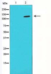
![HIF1 alpha antibody [GT10211] detects HIF1 alpha protein by Western blot analysis. A. 30 μg HepG2 whole cell lysate/extract (untreated) B. 30 μg HepG2 whole cell lysate/extract (500 μM CoCl2 treatment for 24 hr) 7.5 % SDS-PAGE HIF1 alpha antibody [GT10211] (GTX628480) dilution: 1:500](https://www.genetex.com/upload/website/prouct_img/normal/GTX628480/GTX628480_41365_WB_CoCl2_w_23061202_366.webp "HIF1 alpha antibody [GT10211] detects HIF1 alpha protein by Western blot analysis. A. 30 μg HepG2 whole cell lysate/extract (untreated) B. 30 μg HepG2 whole cell lysate/extract (500 μM CoCl2 treatment for 24 hr) 7.5 % SDS-PAGE HIF1 alpha antibody [GT10211] (GTX628480) dilution: 1:500")
![Untreated (–) and treated (+) HeLa whole cell extracts (30 μg) were separated by 5% SDS-PAGE, and the membrane was blotted with HIF1 alpha antibody [GT10211] (GTX628480) diluted at 1:1000. The HRP-conjugated anti-mouse IgG antibody (GTX213111-01) was used to detect the primary antibody.](https://www.genetex.com/upload/website/prouct_img/normal/GTX628480/GTX628480_44503_20211112_WB_treatment_CoCl2_w_23061202_360.webp "Untreated (–) and treated (+) HeLa whole cell extracts (30 μg) were separated by 5% SDS-PAGE, and the membrane was blotted with HIF1 alpha antibody [GT10211] (GTX628480) diluted at 1:1000. The HRP-conjugated anti-mouse IgG antibody (GTX213111-01) was used to detect the primary antibody.")


HIF1 alpha antibody [GT10211] detects HIF1 alpha protein at nucleus by immunofluorescent analysis. Sample: Mock and treated HeLa cells were fixed in 4% paraformaldehyde at RT for 15 min. Green: HIF1 alpha stained by HIF1 alpha antibody [GT10211] (GTX628480) diluted at 1:500. Blue: Fluoroshield with DAPI (GTX30920).
HIF1 alpha antibody [GT10211]
GTX628480
ApplicationsImmunoFluorescence, ImmunoPrecipitation, Western Blot, ImmunoCytoChemistry, ImmunoHistoChemistry, ImmunoHistoChemistry Paraffin
Product group Antibodies
ReactivityHuman, Rat
TargetHIF1A
Overview
- SupplierGeneTex
- Product NameHIF1 alpha antibody [GT10211]
- Delivery Days Customer9
- Application Supplier NoteWB: 1:100-1:3000. ICC/IF: 1:100-1:1000. IP: 1:100-1:500. *Optimal dilutions/concentrations should be determined by the researcher.Not tested in other applications.
- ApplicationsImmunoFluorescence, ImmunoPrecipitation, Western Blot, ImmunoCytoChemistry, ImmunoHistoChemistry, ImmunoHistoChemistry Paraffin
- CertificationResearch Use Only
- ClonalityMonoclonal
- Clone IDGT10211
- Concentration1 mg/ml
- ConjugateUnconjugated
- Gene ID3091
- Target nameHIF1A
- Target descriptionhypoxia inducible factor 1 subunit alpha
- Target synonymsHIF-1-alpha, HIF-1A, HIF-1alpha, HIF1, HIF1-ALPHA, MOP1, PASD8, bHLHe78, hypoxia-inducible factor 1-alpha, ARNT interacting protein, PAS domain-containing protein 8, basic-helix-loop-helix-PAS protein MOP1, class E basic helix-loop-helix protein 78, hypoxia inducible factor 1 alpha subunit, hypoxia inducible factor 1, alpha subunit (basic helix-loop-helix transcription factor), hypoxia-inducible factor1alpha, member of PAS protein 1, member of PAS superfamily 1
- HostMouse
- IsotypeIgG2a
- Protein IDQ16665
- Protein NameHypoxia-inducible factor 1-alpha
- Scientific DescriptionHypoxia-inducible factor-1 (HIF1) is a transcription factor found in mammalian cells cultured under reduced oxygen tension that plays an essential role in cellular and systemic homeostatic responses to hypoxia. HIF1 is a heterodimer composed of an alpha subunit and a beta subunit. The beta subunit has been identified as the aryl hydrocarbon receptor nuclear translocator (ARNT). This gene encodes the alpha subunit of HIF-1. Overexpression of a natural antisense transcript (aHIF) of this gene has been shown to be associated with nonpapillary renal carcinomas. Two alternative transcripts encoding different isoforms have been identified. [provided by RefSeq]
- ReactivityHuman, Rat
- Storage Instruction-20°C or -80°C,2°C to 8°C
- UNSPSC41116161
Datasheet
Related products
Product group Antibodies
HIF1A AntibodyCSB-PA002906
ApplicationsWestern Blot, ELISA, ImmunoHistoChemistry
ReactivityHuman, Mouse, Rat
TargetHIF1A
- SizePrice

Product group Antibodies
Anti-HIF-1 alpha/HIF1A Antibody Picoband(r)A00013-1-CARRIER-FREE
ApplicationsWestern Blot, ELISA
ReactivityHuman
TargetHIF1A
- SizePrice
Product group Antibodies
Anti-HIF1A AntibodyA37506
ApplicationsWestern Blot, ImmunoHistoChemistry
ReactivityHuman, Mouse, Rat
- SizePrice
Product group Antibodies

HIF1A / HIF1 Alpha AntibodyLS-C831817
ApplicationsELISA, ImmunoHistoChemistry
ReactivityHuman, Mouse
TargetHIF1A
- SizePrice

Product group Antibodies
Anti-HIF1A AntibodyHPA000907
ApplicationsImmunoCytoChemistry
ReactivityHuman
TargetHIF1A
- SizePrice
![Untreated (–) and treated (+) HCT116 whole cell extracts (30 μg) were separated by 5% SDS-PAGE, and the membrane was blotted with HIF1 alpha antibody [HL3011] (GTX640424) diluted at 1:1000. The HRP-conjugated anti-rabbit IgG antibody (GTX213110-01) was used to detect the primary antibody.](https://www.genetex.com/upload/website/prouct_img/normal/GTX640424/GTX640424_T-45425_20240614_WB_treatment_hypoxia_24061802_276.webp)
Product group Antibodies
HIF1 alpha antibody [HL3011]GTX640424
ApplicationsImmunoFluorescence, Western Blot, ImmunoCytoChemistry, ImmunoHistoChemistry, ImmunoHistoChemistry Paraffin
ReactivityHuman, Mouse
TargetHIF1A
- SizePrice
![Untreated (–) and treated (+) HeLa whole cell extract (30 μg) were separated by 7.5% SDS-PAGE, and the membrane was blotted with HIF1 alpha antibody [HL3154] (GTX640664) diluted at 1:1000. The HRP-conjugated anti-rabbit IgG antibody (GTX213110-01) was used to detect the primary antibody.](https://www.genetex.com/upload/website/prouct_img/normal/GTX640664/GTX640664_T-45481_20240726_WB_treatment_CoCl2_24080622_872.webp)
Product group Antibodies
HIF1 alpha antibody [HL3154]GTX640664
ApplicationsImmunoFluorescence, Western Blot, ImmunoCytoChemistry, ImmunoHistoChemistry, ImmunoHistoChemistry Paraffin
ReactivityHuman, Mouse, Rat
TargetHIF1A
- SizePrice
Product group Antibodies
HIF1 alpha antibodyGTX127309
ApplicationsImmunoFluorescence, ImmunoPrecipitation, Western Blot, ChIP Chromatin ImmunoPrecipitation, ImmunoCytoChemistry, ImmunoHistoChemistry, ImmunoHistoChemistry Frozen, ImmunoHistoChemistry Paraffin
ReactivityBovine, Human, Mouse, Rabbit, Rat
TargetHIF1A
- SizePrice
![Untreated (–) and treated (+) NIH-3T3 whole cell extracts (30 μg) were separated by 5% SDS-PAGE, and the membrane was blotted with HIF1 alpha antibody [GT122] (GTX629766) diluted at 1:1000. The HRP-conjugated anti-mouse IgG antibody (GTX213111-01) was used to detect the primary antibody, and the signal was developed with Trident ECL plus-Enhanced.](https://www.genetex.com/upload/website/prouct_img/normal/GTX629766/GTX629766_44615_20220318_WB_M_treatment_CoCl2_w_23051500_576.webp)
Product group Antibodies
HIF1 alpha antibody [GT122]GTX629766
ApplicationsImmunoFluorescence, Western Blot, ImmunoCytoChemistry
ReactivityHuman, Mouse
TargetHIF1A
- SizePrice