

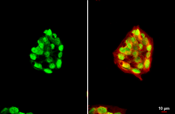
HNF1 alpha antibody [N1N3] detects HNF1 alpha protein at nucleus by immunofluorescent analysis. Sample: HepG2 cells were fixed in 4% paraformaldehyde at RT for 15 min. Green: HNF1 alpha stained by HNF1 alpha antibody [N1N3] (GTX113850) diluted at 1:500. Red: alpha Tubulin, a cytoskeleton marker, stained by alpha Tubulin antibody [GT114] (GTX628802) diluted at 1:1000. Scale bar= 10μm.
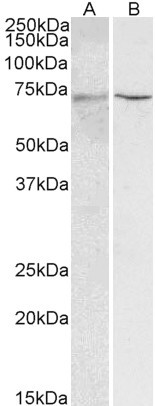
![Mouse tissue extract (50 μg) was separated by 7.5% SDS-PAGE, and the membrane was blotted with HNF1 alpha antibody [N1N3] (GTX113850) diluted at 1:2000. The HRP-conjugated anti-rabbit IgG antibody (GTX213110-01) was used to detect the primary antibody.](https://www.genetex.com/upload/website/prouct_img/normal/GTX113850/GTX113850_44665_20220429_WB_M_liver_22071823_768.webp "Mouse tissue extract (50 μg) was separated by 7.5% SDS-PAGE, and the membrane was blotted with HNF1 alpha antibody [N1N3] (GTX113850) diluted at 1:2000. The HRP-conjugated anti-rabbit IgG antibody (GTX213110-01) was used to detect the primary antibody.")
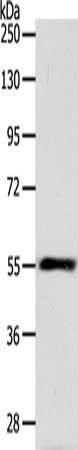
![Various whole cell extracts (30 μg) were separated by 7.5% SDS-PAGE, and the membrane was blotted with HNF1 alpha antibody [N1N3] (GTX113850) diluted at 1:2000. The HRP-conjugated anti-rabbit IgG antibody (GTX213110-01) was used to detect the primary antibody.](https://www.genetex.com/upload/website/prouct_img/normal/GTX113850/GTX113850_44665_20220429_WB_22071823_792.webp "Various whole cell extracts (30 μg) were separated by 7.5% SDS-PAGE, and the membrane was blotted with HNF1 alpha antibody [N1N3] (GTX113850) diluted at 1:2000. The HRP-conjugated anti-rabbit IgG antibody (GTX213110-01) was used to detect the primary antibody.")
![Various whole cell extracts (30 μg) were separated by 7.5% SDS-PAGE, and the membrane was blotted with HNF1 alpha antibody [N1N3] (GTX113850) diluted at 1:2000. The HRP-conjugated anti-rabbit IgG antibody (GTX213110-01) was used to detect the primary antibody. Corresponding RNA expression data for the same cell lines are based on Human Protein Atlas program.](https://www.genetex.com/upload/website/prouct_img/normal/GTX113850/GTX113850_43908_20200410_WB_TPM_watermark_w_23060501_136.webp "Various whole cell extracts (30 μg) were separated by 7.5% SDS-PAGE, and the membrane was blotted with HNF1 alpha antibody [N1N3] (GTX113850) diluted at 1:2000. The HRP-conjugated anti-rabbit IgG antibody (GTX213110-01) was used to detect the primary antibody. Corresponding RNA expression data for the same cell lines are based on Human Protein Atlas program.")
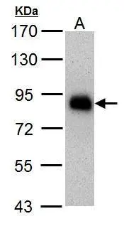
![Immunoprecipitation of HNF1 alpha protein from HepG2 whole cell extracts using 5 μg of HNF1 alpha antibody [N1N3] (GTX113850). Western blot analysis was performed using HNF1 alpha antibody [N1N3] (GTX113850). EasyBlot anti-Rabbit IgG (GTX221666-01) was used as a secondary reagent.](https://www.genetex.com/upload/website/prouct_img/normal/GTX113850/GTX113850_41080_20150317_IP_w_23060501_859.webp "Immunoprecipitation of HNF1 alpha protein from HepG2 whole cell extracts using 5 μg of HNF1 alpha antibody [N1N3] (GTX113850). Western blot analysis was performed using HNF1 alpha antibody [N1N3] (GTX113850). EasyBlot anti-Rabbit IgG (GTX221666-01) was used as a secondary reagent.")
HNF1 alpha antibody [N1N3] detects HNF1 alpha protein at nucleus by immunofluorescent analysis. Sample: HepG2 cells were fixed in 4% paraformaldehyde at RT for 15 min. Green: HNF1 alpha stained by HNF1 alpha antibody [N1N3] (GTX113850) diluted at 1:500. Red: alpha Tubulin, a cytoskeleton marker, stained by alpha Tubulin antibody [GT114] (GTX628802) diluted at 1:1000. Scale bar= 10μm.
HNF1 alpha antibody [N1N3]
GTX113850
ApplicationsImmunoFluorescence, ImmunoPrecipitation, Western Blot, ImmunoCytoChemistry
Product group Antibodies
ReactivityHuman, Mouse
TargetHNF1A
Overview
- SupplierGeneTex
- Product NameHNF1 alpha antibody [N1N3]
- Delivery Days Customer9
- Application Supplier NoteWB: 1:1000-1:10000. ICC/IF: 1:100-1:1000. IP: 1:100-1:500. *Optimal dilutions/concentrations should be determined by the researcher.Not tested in other applications.
- ApplicationsImmunoFluorescence, ImmunoPrecipitation, Western Blot, ImmunoCytoChemistry
- CertificationResearch Use Only
- ClonalityPolyclonal
- Concentration1.27 mg/ml
- ConjugateUnconjugated
- Gene ID6927
- Target nameHNF1A
- Target descriptionHNF1 homeobox A
- Target synonymsHNF-1-alpha, HNF-1A, HNF1, HNF1alpha, HNF4A, IDDM20, LFB1, MODY3, TCF-1, TCF1, hepatocyte nuclear factor 1-alpha, albumin proximal factor, hepatic nuclear factor 1, interferon production regulator factor, liver-specific transcription factor LF-B1, transcription factor 1, hepatic
- HostRabbit
- IsotypeIgG
- Protein IDP20823
- Protein NameHepatocyte nuclear factor 1-alpha
- Scientific DescriptionThe protein encoded by this gene is a transcription factor required for the expression of several liver-specific genes. The encoded protein functions as a homodimer and binds to the inverted palindrome 5-GTTAATNATTAAC-3. Defects in this gene are a cause of maturity onset diabetes of the young type 3 (MODY3) and also can result in the appearance of hepatic adenomas. [provided by RefSeq]
- ReactivityHuman, Mouse
- Storage Instruction-20°C or -80°C,2°C to 8°C
- UNSPSC41116161
Datasheet
Related products
Product group Antibodies
Anti-HNF1 alpha Antibody107-11093
ApplicationsWestern Blot, ImmunoHistoChemistry, ImmunoHistoChemistry Paraffin
ReactivityHuman
TargetHNF1A
- SizePrice
Product group Antibodies
Anti-HNF1 alpha AntibodyA326253
ApplicationsImmunoFluorescence, Western Blot, ELISA, ImmunoHistoChemistry
ReactivityHuman, Mouse, Rat
- SizePrice
Product group Antibodies

HNF1A / HNF1 AntibodyLS-C831045
ApplicationsELISA, ImmunoHistoChemistry
ReactivityHuman, Mouse, Rat
TargetHNF1A
- SizePrice
Product group Antibodies
HNF1A Polyclonal AntibodyBS-25295R
ApplicationsImmunoFluorescence, ImmunoHistoChemistry, ImmunoHistoChemistry Frozen, ImmunoHistoChemistry Paraffin
ReactivityBovine, Human, Mouse, Porcine, Rat, Sheep
TargetHNF1A
- SizePrice
Product group Antibodies
HNF1A AntibodyCSB-PA944576
ApplicationsWestern Blot, ELISA
ReactivityHuman, Mouse, Rat
TargetHNF1A
- SizePrice
Product group Antibodies

Goat anti-HNF1AEB06701
ApplicationsImmunoFluorescence, Western Blot, ELISA, ImmunoHistoChemistry
ReactivityBovine, Canine, Human, Mouse, Porcine, Rat
TargetHNF1A
- SizePrice
Product group Antibodies
ApplicationsImmunoPrecipitation, Western Blot, ImmunoCytoChemistry, ImmunoHistoChemistry
TargetHNF1A
- SizePrice
![Various whole cell extracts (30 μg) were separated by 7.5% SDS-PAGE, and the membrane was blotted with HNF1 alpha antibody [HL3229] (GTX640870) diluted at 1:1000. The HRP-conjugated anti-rabbit IgG antibody (GTX213110-01) was used to detect the primary antibody. Corresponding RNA expression data for the same cell lines are based on Human Protein Atlas program.](https://www.genetex.com/upload/website/prouct_img/normal/GTX640870/GTX640870_T-45509_20240906_WB_TPM_watermark_24091102_352.webp)
Product group Antibodies
HNF1 alpha antibody [HL3229]GTX640870
ApplicationsImmunoFluorescence, Western Blot, ImmunoCytoChemistry, ImmunoHistoChemistry, ImmunoHistoChemistry Paraffin
ReactivityHuman, Mouse
TargetHNF1A
- SizePrice
Product group Antibodies
HNF1 alpha antibody [GT4110]GTX628240
ApplicationsWestern Blot, ImmunoHistoChemistry, ImmunoHistoChemistry Paraffin
ReactivityHuman, Mouse
TargetHNF1A
- SizePrice
Product group Antibodies
Anti-HNF1A AntibodyHPA035231
ApplicationsWestern Blot, ImmunoCytoChemistry, ImmunoHistoChemistry
ReactivityHuman
TargetHNF1A
- SizePrice