

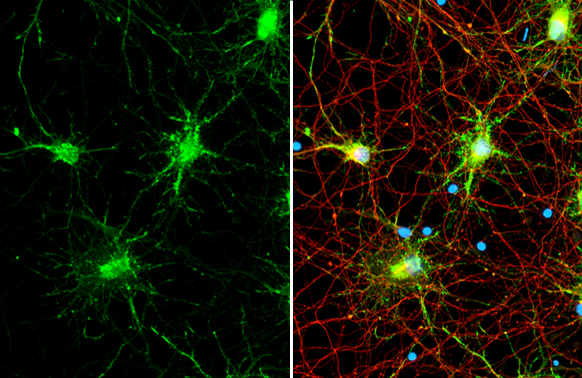
iNOS antibody [HL1213] detects iNOS protein by immunofluorescent analysis. Sample: DIV9 rat cortical neuron and Glia cell cells were fixed in 4% paraformaldehyde at RT for 15 min. Green: iNOS stained by iNOS antibody [HL1213] (GTX636531) diluted at 1:2500. Red: Tau, an axon marker, stained by Tau antibody [GT287] (GTX634809) diluted at 1:500. Blue: Fluoroshield with DAPI (GTX30920).
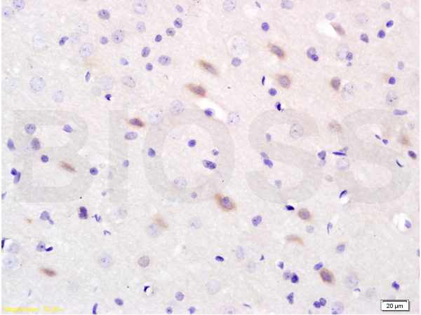
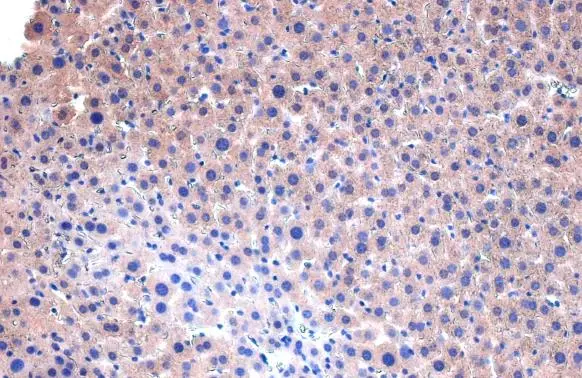
![iNOS antibody [HL1213] detects iNOS protein at cytoplasm by immunohistochemical analysis. Sample: Paraffin-embedded mouse liver. iNOS stained by iNOS antibody [HL1213] (GTX636531) diluted at 1:50. Antigen Retrieval: Citrate buffer, pH 6.0, 15 min](https://www.genetex.com/upload/website/prouct_img/normal/GTX636531/GTX636531_T-44473_20211224_IHC-P_M_w_23061202_895.webp "iNOS antibody [HL1213] detects iNOS protein at cytoplasm by immunohistochemical analysis. Sample: Paraffin-embedded mouse liver. iNOS stained by iNOS antibody [HL1213] (GTX636531) diluted at 1:50. Antigen Retrieval: Citrate buffer, pH 6.0, 15 min")
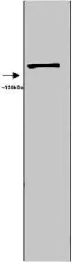
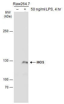
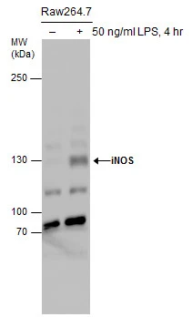
![Untreated (–) and treated (+) Raw264.7 whole cell extracts (30 μg) were separated by 5% SDS-PAGE, and the membrane was blotted with iNOS antibody [HL1213] (GTX636531) diluted at 1:1000. The HRP-conjugated anti-rabbit IgG antibody (GTX213110-01) was used to detect the primary antibody.](https://www.genetex.com/upload/website/prouct_img/normal/GTX636531/GTX636531_45278_20240112_WB_M_treatment_LPS_25041720_757.webp "Untreated (–) and treated (+) Raw264.7 whole cell extracts (30 μg) were separated by 5% SDS-PAGE, and the membrane was blotted with iNOS antibody [HL1213] (GTX636531) diluted at 1:1000. The HRP-conjugated anti-rabbit IgG antibody (GTX213110-01) was used to detect the primary antibody.")
iNOS antibody [HL1213] detects iNOS protein by immunofluorescent analysis. Sample: DIV9 rat cortical neuron and Glia cell cells were fixed in 4% paraformaldehyde at RT for 15 min. Green: iNOS stained by iNOS antibody [HL1213] (GTX636531) diluted at 1:2500. Red: Tau, an axon marker, stained by Tau antibody [GT287] (GTX634809) diluted at 1:500. Blue: Fluoroshield with DAPI (GTX30920).
iNOS antibody [HL1213]
GTX636531
ApplicationsImmunoFluorescence, Western Blot, ImmunoCytoChemistry, ImmunoHistoChemistry, ImmunoHistoChemistry Paraffin
Product group Antibodies
ReactivityMouse, Rat
TargetNos2
Overview
- SupplierGeneTex
- Product NameiNOS antibody [HL1213]
- Delivery Days Customer9
- Application Supplier NoteWB: 1:500-1:3000. ICC/IF: 1:100-1:2500. *Optimal dilutions/concentrations should be determined by the researcher.Not tested in other applications.
- ApplicationsImmunoFluorescence, Western Blot, ImmunoCytoChemistry, ImmunoHistoChemistry, ImmunoHistoChemistry Paraffin
- CertificationResearch Use Only
- ClonalityMonoclonal
- Clone IDHL1213
- Concentration1 mg/ml
- ConjugateUnconjugated
- Gene ID18126
- Target nameNos2
- Target descriptionnitric oxide synthase 2, inducible
- Target synonymsMAC-NOS, NOS-II, Nos-2, Nos2a, i-NOS, iNOS, nitric oxide synthase, inducible, NOS type II, inducible NO synthase, inducible NOS, inducible nitric oxide synthase, macrophage NOS, nitric oxide synthase 2, inducible, macrophage, peptidyl-cysteine S-nitrosylase NOS2
- HostRabbit
- IsotypeIgG
- Protein IDP29477
- Protein NameNitric oxide synthase, inducible
- Scientific DescriptionNitric oxide is a reactive free radical which acts as a biologic mediator in several processes, including neurotransmission and antimicrobial and antitumoral activities. This gene encodes a nitric oxide synthase that is inducible by a combination of lipopolysaccharide and certain cytokines. Three transcript variants encoding two different isoforms have been found for this gene. [provided by RefSeq, Sep 2015]
- ReactivityMouse, Rat
- Storage Instruction-20°C or -80°C,2°C to 8°C
- UNSPSC41116161
Datasheet
Related products

Product group Antibodies
Anti-iNOS/Nos2 Antibody Picoband(r)A00368-4-CARRIER-FREE
ApplicationsFlow Cytometry, Western Blot, ELISA
ReactivityMouse
TargetNos2
- SizePrice
Product group Antibodies
References
iNOS Polyclonal AntibodyBS-2072R
ApplicationsFlow Cytometry, ImmunoFluorescence, Western Blot, ELISA, ImmunoCytoChemistry, ImmunoHistoChemistry, ImmunoHistoChemistry Frozen, ImmunoHistoChemistry Paraffin
ReactivityMouse, Rabbit, Rat
TargetNos2
- SizePrice
Product group Antibodies
ApplicationsImmunoPrecipitation, Western Blot, ImmunoCytoChemistry, ImmunoHistoChemistry
ReactivityMouse
TargetNos2
- SizePrice
Product group Antibodies
References
iNOS antibodyGTX17504
ApplicationsImmunoFluorescence, Western Blot, ImmunoCytoChemistry, ImmunoHistoChemistry, ImmunoHistoChemistry Frozen, ImmunoHistoChemistry Paraffin, Other Application
ReactivityAmphibian, Avian, Feline, Fish, Human, Mouse, Porcine, Rabbit, Rat
TargetNos2
- SizePrice
Product group Antibodies
iNOS antibodyGTX74171
ApplicationsImmunoFluorescence, Western Blot, ImmunoCytoChemistry, ImmunoHistoChemistry, ImmunoHistoChemistry Frozen, ImmunoHistoChemistry Paraffin
ReactivityMouse, Rat
TargetNos2
- SizePrice
Product group Antibodies
iNOS antibodyGTX127952
ApplicationsWestern Blot
ReactivityMouse
TargetNos2
- SizePrice
Product group Antibodies
iNOS antibodyGTX130182
ApplicationsImmunoFluorescence, Western Blot, ImmunoCytoChemistry
ReactivityMouse, Rat
TargetNos2
- SizePrice
Product group Antibodies
iNOS antibodyGTX130246
ApplicationsImmunoFluorescence, Western Blot, ImmunoCytoChemistry, ImmunoHistoChemistry, ImmunoHistoChemistry Paraffin
ReactivityMouse, Rat
TargetNos2
- SizePrice