

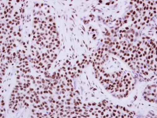
Immunohistochemical analysis of paraffin-embedded human breast cancer, using KAP1(GTX102226) antibody at 1:500 dilution.
Antigen Retrieval: Trilogy? (EDTA based, pH 8.0) buffer, 15min
![KAP1 antibody [N3C2], Internal detects KAP1 protein at nucleus by immunohistochemical analysis. Sample: Paraffin-embedded mouse colon. KAP1 stained by KAP1 antibody [N3C2], Internal (GTX102226) diluted at 1:500. Antigen Retrieval: Citrate buffer, pH 6.0, 15 min](https://www.genetex.com/upload/website/prouct_img/normal/GTX102226/GTX102226_44490_20211210_IHC-P_M_1_w_23060100_290.webp "KAP1 antibody [N3C2], Internal detects KAP1 protein at nucleus by immunohistochemical analysis. Sample: Paraffin-embedded mouse colon. KAP1 stained by KAP1 antibody [N3C2], Internal (GTX102226) diluted at 1:500. Antigen Retrieval: Citrate buffer, pH 6.0, 15 min")
")
![KAP1 antibody [N3C2], Internal detects KAP1 protein at nucleus by immunohistochemical analysis. Sample: Paraffin-embedded mouse intestine. KAP1 stained by KAP1 antibody [N3C2], Internal (GTX102226) diluted at 1:500. Antigen Retrieval: Citrate buffer, pH 6.0, 15 min](https://www.genetex.com/upload/website/prouct_img/normal/GTX102226/GTX102226_44490_20211210_IHC-P_M_w_23060100_818.webp "KAP1 antibody [N3C2], Internal detects KAP1 protein at nucleus by immunohistochemical analysis. Sample: Paraffin-embedded mouse intestine. KAP1 stained by KAP1 antibody [N3C2], Internal (GTX102226) diluted at 1:500. Antigen Retrieval: Citrate buffer, pH 6.0, 15 min")
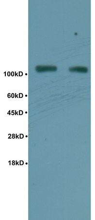
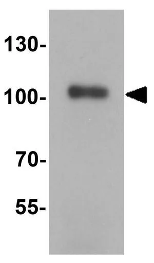
 7.5% SDS-PAGE The immunoprecipitated KAP1 protein was detected by KAP1 antibody (GTX102226) diluted at 1:1000. EasyBlot anti-rabbit IgG (GTX221666-01) was used as a secondary reagent.")
 A:NIH-3T3 7.5% SDS PAGE GTX102226 diluted at 1:1000 The HRP-conjugated anti-rabbit IgG antibody (GTX213110-01) was used to detect the primary antibody.")
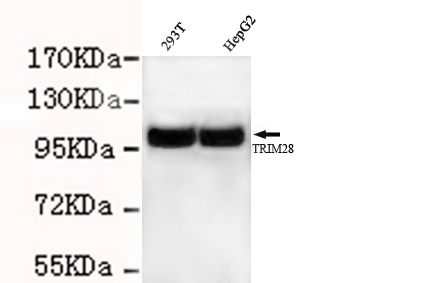
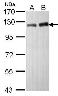
![Various whole cell extracts (30 μg) were separated by 5% SDS-PAGE, and the membrane was blotted with KAP1 antibody [N3C2], Internal (GTX102226) diluted at 1:10000. The HRP-conjugated anti-rabbit IgG antibody (GTX213110-01) was used to detect the primary antibody.](https://www.genetex.com/upload/website/prouct_img/normal/GTX102226/GTX102226_44489_20211112_WB_w_23060100_614.webp "Various whole cell extracts (30 μg) were separated by 5% SDS-PAGE, and the membrane was blotted with KAP1 antibody [N3C2], Internal (GTX102226) diluted at 1:10000. The HRP-conjugated anti-rabbit IgG antibody (GTX213110-01) was used to detect the primary antibody.")
![KAP1 antibody [N3C2], Internal detects KAP1 protein at nucleus by immunofluorescent analysis. Sample: HeLa cells were fixed in 4% paraformaldehyde at RT for 15 min. Green: KAP1 stained by KAP1 antibody [N3C2], Internal (GTX102226) diluted at 1:2000. Red: phalloidin, a cytoskeleton marker, diluted at 1:200. Scale bar= 10 μm.](https://www.genetex.com/upload/website/prouct_img/normal/GTX102226/GTX102226_43628_20200219_ICC_IF_w_23060100_583.webp "KAP1 antibody [N3C2], Internal detects KAP1 protein at nucleus by immunofluorescent analysis. Sample: HeLa cells were fixed in 4% paraformaldehyde at RT for 15 min. Green: KAP1 stained by KAP1 antibody [N3C2], Internal (GTX102226) diluted at 1:2000. Red: phalloidin, a cytoskeleton marker, diluted at 1:200. Scale bar= 10 μm.")

Immunohistochemical analysis of paraffin-embedded human breast cancer, using KAP1(GTX102226) antibody at 1:500 dilution.
Antigen Retrieval: Trilogy? (EDTA based, pH 8.0) buffer, 15min
KAP1 antibody [N3C2], Internal
GTX102226
ApplicationsImmunoFluorescence, ImmunoPrecipitation, Western Blot, ChIP Chromatin ImmunoPrecipitation, ImmunoCytoChemistry, ImmunoHistoChemistry, ImmunoHistoChemistry Paraffin
Product group Antibodies
ReactivityHuman, Mouse
TargetTRIM28
Overview
- SupplierGeneTex
- Product NameKAP1 antibody [N3C2], Internal
- Delivery Days Customer9
- Application Supplier NoteWB: 1:1000-1:10000. ICC/IF: 1:100-1:1000. IHC-P: 1:100-1:1000. IP: 1:500-1:1000. *Optimal dilutions/concentrations should be determined by the researcher.Not tested in other applications.
- ApplicationsImmunoFluorescence, ImmunoPrecipitation, Western Blot, ChIP Chromatin ImmunoPrecipitation, ImmunoCytoChemistry, ImmunoHistoChemistry, ImmunoHistoChemistry Paraffin
- CertificationResearch Use Only
- ClonalityPolyclonal
- Concentration0.48 mg/ml
- ConjugateUnconjugated
- Gene ID10155
- Target nameTRIM28
- Target descriptiontripartite motif containing 28
- Target synonymsKAP1, PPP1R157, RNF96, TF1B, TIF1B, TIF1beta, WT7, transcription intermediary factor 1-beta, E3 SUMO-protein ligase TRIM28, KAP-1, KRAB [Kruppel-associated box domain]-associated protein 1, KRAB-interacting protein 1, KRIP-1, RING finger protein 96, RING-type E3 ubiquitin transferase TIF1-beta, TIF1-beta, nuclear corepressor KAP-1, protein phosphatase 1, regulatory subunit 157, transcriptional intermediary factor 1-beta
- HostRabbit
- IsotypeIgG
- Protein IDQ13263
- Protein NameTranscription intermediary factor 1-beta
- Scientific DescriptionThe protein encoded by this gene mediates transcriptional control by interaction with the Kruppel-associated box repression domain found in many transcription factors. The protein localizes to the nucleus and is thought to associate with specific chromatin regions. The protein is a member of the tripartite motif family. This tripartite motif includes three zinc-binding domains, a RING, a B-box type 1 and a B-box type 2, and a coiled-coil region. [provided by RefSeq]
- ReactivityHuman, Mouse
- Storage Instruction-20°C or -80°C,2°C to 8°C
- UNSPSC41116161
Datasheet
Related products
Product group Antibodies
Anti-TRIM28 AntibodyA35695
ApplicationsWestern Blot, ImmunoCytoChemistry, ImmunoHistoChemistry
ReactivityCanine, Human, Mouse, Rat
- SizePrice
Product group Antibodies

Anti-TRIM28 [RAB-C429]AB01909-1.1-BT
ApplicationsImmunoFluorescence, ImmunoPrecipitation
ReactivityHuman
TargetTRIM28
- SizePrice
Product group Antibodies
Anti-TRIM28 Antibody144-02245
ApplicationsWestern Blot, ImmunoHistoChemistry
ReactivityHuman, Mouse, Rat
TargetTRIM28
- SizePrice
Product group Antibodies
References
ApplicationsFlow Cytometry, ImmunoFluorescence, Western Blot, ELISA, ImmunoCytoChemistry, ImmunoHistoChemistry, ImmunoHistoChemistry Frozen, ImmunoHistoChemistry Paraffin
ReactivityHuman, Mouse, Rat
TargetTRIM28
- SizePrice
Product group Antibodies
References

Goat anti-KAP1 / TRIM28EB05810
ApplicationsFlow Cytometry, ImmunoFluorescence, ELISA, ImmunoHistoChemistry
ReactivityHuman, Mouse
TargetTRIM28
- SizePrice
Product group Antibodies
Trim28 Polyclonal AntibodyCAC07189
ApplicationsImmunoPrecipitation, Western Blot, ELISA, ImmunoHistoChemistry
TargetTRIM28
- SizePrice
Product group Antibodies
TRIM28 AntibodyCSB-PA05254A0RB
ApplicationsImmunoPrecipitation, Western Blot, ELISA, ImmunoHistoChemistry
ReactivityHuman
TargetTRIM28
- SizePrice
Product group Antibodies
KAP1 antibodyGTX31274
ApplicationsWestern Blot, ELISA
ReactivityHuman, Mouse, Rat
TargetTRIM28
- SizePrice
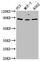
![WB analysis of various samples using GTX08968 KAP1 antibody [GT1161]. Dilution : 1:1000 Loading : 25 μg](https://www.genetex.com/upload/website/prouct_img/normal/GTX08968/GTX08968_20200508_WB_w_23053123_747.webp)
Product group Antibodies
KAP1 antibody [GT1161]GTX08968
ApplicationsImmunoPrecipitation, Western Blot, ImmunoHistoChemistry, ImmunoHistoChemistry Paraffin
ReactivityHuman, Mouse, Rat
TargetTRIM28
- SizePrice
Product group Antibodies
KAP1 antibody [N1N2], N-termGTX102227
ApplicationsImmunoFluorescence, Western Blot, ImmunoCytoChemistry, ImmunoHistoChemistry, ImmunoHistoChemistry Paraffin
ReactivityHuman, Mouse
TargetTRIM28
- SizePrice