

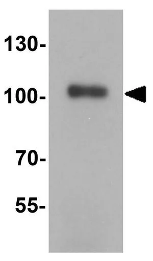
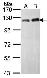
WB analysis of human testis tissue lysate using GTX31274 KAP1 antibody. Working concentration : 1 μg/ml
WB analysis of human testis tissue lysate using GTX31274 KAP1 antibody. Working concentration : 1 μg/ml
KAP1 antibody
GTX31274
ApplicationsWestern Blot, ELISA
Product group Antibodies
ReactivityHuman, Mouse, Rat
TargetTRIM28
Overview
- SupplierGeneTex
- Product NameKAP1 antibody
- Delivery Days Customer9
- Application Supplier NoteWB: 1 - 2 microg/mL. *Optimal dilutions/concentrations should be determined by the researcher.Not tested in other applications.
- ApplicationsWestern Blot, ELISA
- CertificationResearch Use Only
- ClonalityPolyclonal
- Concentration1 mg/ml
- ConjugateUnconjugated
- Gene ID10155
- Target nameTRIM28
- Target descriptiontripartite motif containing 28
- Target synonymsKAP1, PPP1R157, RNF96, TF1B, TIF1B, TIF1beta, WT7, transcription intermediary factor 1-beta, E3 SUMO-protein ligase TRIM28, KAP-1, KRAB [Kruppel-associated box domain]-associated protein 1, KRAB-interacting protein 1, KRIP-1, RING finger protein 96, RING-type E3 ubiquitin transferase TIF1-beta, TIF1-beta, nuclear corepressor KAP-1, protein phosphatase 1, regulatory subunit 157, transcriptional intermediary factor 1-beta
- HostRabbit
- IsotypeIgG
- Protein IDQ13263
- Protein NameTranscription intermediary factor 1-beta
- Scientific DescriptionThe protein encoded by this gene mediates transcriptional control by interaction with the Kruppel-associated box repression domain found in many transcription factors. The protein localizes to the nucleus and is thought to associate with specific chromatin regions. The protein is a member of the tripartite motif family. This tripartite motif includes three zinc-binding domains, a RING, a B-box type 1 and a B-box type 2, and a coiled-coil region. [provided by RefSeq, Jul 2008]
- ReactivityHuman, Mouse, Rat
- Storage Instruction-20°C or -80°C,2°C to 8°C
- UNSPSC41116161
Datasheet
Related products
Product group Antibodies
Anti-TRIM28 AntibodyA35695
ApplicationsWestern Blot, ImmunoCytoChemistry, ImmunoHistoChemistry
ReactivityCanine, Human, Mouse, Rat
- SizePrice
Product group Antibodies

Anti-TRIM28 [RAB-C429]AB01909-1.1-BT
ApplicationsImmunoFluorescence, ImmunoPrecipitation
ReactivityHuman
TargetTRIM28
- SizePrice
Product group Antibodies
Anti-TRIM28 Antibody144-02245
ApplicationsWestern Blot, ImmunoHistoChemistry
ReactivityHuman, Mouse, Rat
TargetTRIM28
- SizePrice
Product group Antibodies
References
ApplicationsFlow Cytometry, ImmunoFluorescence, Western Blot, ELISA, ImmunoCytoChemistry, ImmunoHistoChemistry, ImmunoHistoChemistry Frozen, ImmunoHistoChemistry Paraffin
ReactivityHuman, Mouse, Rat
TargetTRIM28
- SizePrice
Product group Antibodies
References

Goat anti-KAP1 / TRIM28EB05810
ApplicationsFlow Cytometry, ImmunoFluorescence, ELISA, ImmunoHistoChemistry
ReactivityHuman, Mouse
TargetTRIM28
- SizePrice
Product group Antibodies
Trim28 Polyclonal AntibodyCAC07189
ApplicationsImmunoPrecipitation, Western Blot, ELISA, ImmunoHistoChemistry
TargetTRIM28
- SizePrice
Product group Antibodies
TRIM28 AntibodyCSB-PA05254A0RB
ApplicationsImmunoPrecipitation, Western Blot, ELISA, ImmunoHistoChemistry
ReactivityHuman
TargetTRIM28
- SizePrice
![WB analysis of various samples using GTX08968 KAP1 antibody [GT1161]. Dilution : 1:1000 Loading : 25 μg](https://www.genetex.com/upload/website/prouct_img/normal/GTX08968/GTX08968_20200508_WB_w_23053123_747.webp)
Product group Antibodies
KAP1 antibody [GT1161]GTX08968
ApplicationsImmunoPrecipitation, Western Blot, ImmunoHistoChemistry, ImmunoHistoChemistry Paraffin
ReactivityHuman, Mouse, Rat
TargetTRIM28
- SizePrice
Product group Antibodies
KAP1 antibody [N3C2], InternalGTX102226
ApplicationsImmunoFluorescence, ImmunoPrecipitation, Western Blot, ChIP Chromatin ImmunoPrecipitation, ImmunoCytoChemistry, ImmunoHistoChemistry, ImmunoHistoChemistry Paraffin
ReactivityHuman, Mouse
TargetTRIM28
- SizePrice
Product group Antibodies
KAP1 antibody [N1N2], N-termGTX102227
ApplicationsImmunoFluorescence, Western Blot, ImmunoCytoChemistry, ImmunoHistoChemistry, ImmunoHistoChemistry Paraffin
ReactivityHuman, Mouse
TargetTRIM28
- SizePrice