

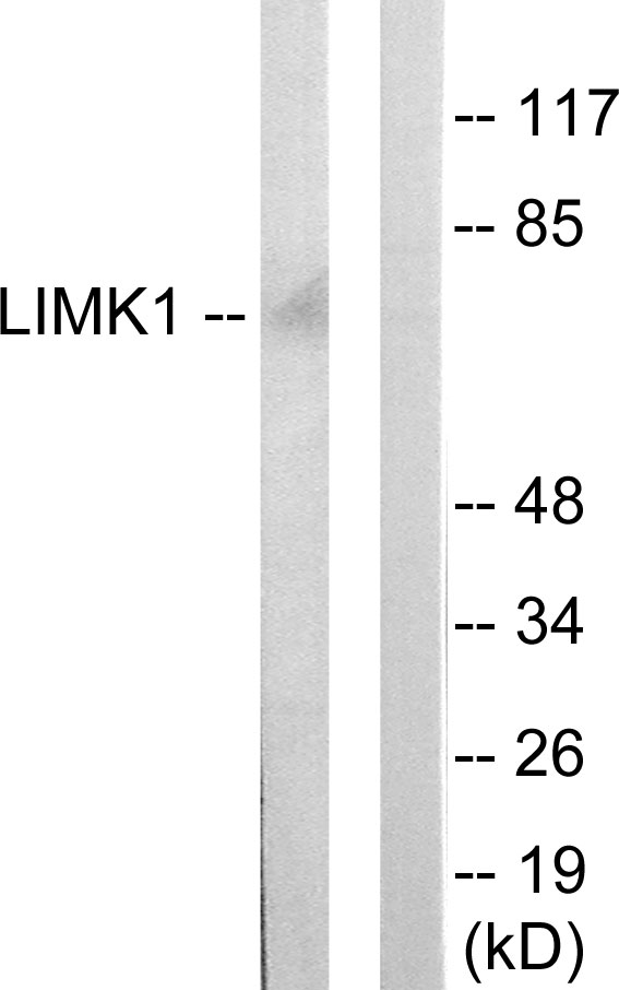
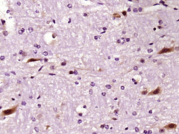
LIM Kinase 1 Antibody
ABX033701
ApplicationsWestern Blot, ELISA, ImmunoHistoChemistry
Product group Antibodies
Overview
- SupplierAbbexa
- Product NameLIM Kinase 1 Antibody
- Delivery Days Customer12
- ApplicationsWestern Blot, ELISA, ImmunoHistoChemistry
- CertificationResearch Use Only
- ClonalityPolyclonal
- ConjugateUnconjugated
- HostRabbit
- UNSPSC12352203
Related products
Product group Antibodies
Anti-LIMK1 AntibodyA97426
ApplicationsWestern Blot, ELISA
ReactivityHuman, Mouse, Rat
- SizePrice
Product group Antibodies
Anti-LIMK1 Antibody144-64575
ApplicationsWestern Blot
ReactivityHuman, Mouse, Rat
TargetLIMK1
- SizePrice
Product group Antibodies
References
LIMK1 Polyclonal AntibodyBS-2775R
ApplicationsImmunoFluorescence, Western Blot, ELISA, ImmunoCytoChemistry, ImmunoHistoChemistry, ImmunoHistoChemistry Frozen, ImmunoHistoChemistry Paraffin
ReactivityBovine, Human, Mouse, Rabbit, Rat
TargetLIMK1
- SizePrice
Product group Antibodies
LIMK1 AntibodyCSB-PA003150
ApplicationsImmunoFluorescence, Western Blot, ELISA, ImmunoHistoChemistry
ReactivityHuman, Mouse, Rat
TargetLIMK1
- SizePrice
Product group Antibodies

LIMK1 / LIMK AntibodyLS-C403296
ApplicationsELISA, ImmunoHistoChemistry
ReactivityHuman, Mouse, Rat
TargetLIMK1
- SizePrice

Product group Antibodies
Anti-LIMK1 AntibodyHPA028064
ApplicationsWestern Blot, ImmunoHistoChemistry
ReactivityHuman
TargetLIMK1
- SizePrice
Product group Antibodies
References
LIMK1 antibodyGTX10561
ApplicationsImmunoFluorescence, Western Blot, ELISA, ImmunoCytoChemistry
ReactivityHuman, Mouse
TargetLIMK1
- SizePrice

Product group Antibodies
Anti-LIMK1 Antibody Picoband(r)PB9716-CARRIER-FREE
ApplicationsWestern Blot
ReactivityHuman, Mouse, Rat
TargetLIMK1
- SizePrice