


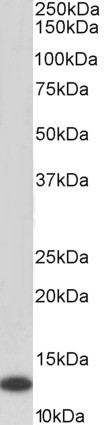
WB analysis of HL-60 lysate using MIF antibody at a dilution of 1:1,000.
WB analysis of HL-60 lysate using MIF antibody at a dilution of 1:1,000.
MIF antibody [4E4]
GTX53741
ApplicationsFlow Cytometry, ImmunoFluorescence, Western Blot, ELISA, ImmunoCytoChemistry
Product group Antibodies
ReactivityHuman
TargetMIF
Overview
- SupplierGeneTex
- Product NameMIF antibody [4E4]
- Delivery Days Customer9
- Application Supplier NoteThe antibody has been tested by ELISA and Western blot analysis to assure specificity and reactivity. Since application varies, however, each investigation should be titrated by the reagent to obtain optimal results. Recommended dilution range for Western blot analysis is 1:500 ~ 2,000. Recommended starting dilution is 1:1,000.
- ApplicationsFlow Cytometry, ImmunoFluorescence, Western Blot, ELISA, ImmunoCytoChemistry
- CertificationResearch Use Only
- ClonalityMonoclonal
- Concentration1 mg/ml
- ConjugateUnconjugated
- Gene ID4282
- Target nameMIF
- Target descriptionmacrophage migration inhibitory factor
- Target synonymsGIF, GLIF, MMIF, macrophage migration inhibitory factor, L-dopachrome isomerase, L-dopachrome tautomerase, epididymis secretory sperm binding protein, macrophage migration inhibitory factor (glycosylation-inhibiting factor), phenylpyruvate tautomerase
- HostMouse
- IsotypeIgG1
- Protein IDP14174
- Protein NameMacrophage migration inhibitory factor
- Scientific DescriptionThis gene encodes a lymphokine involved in cell-mediated immunity, immunoregulation, and inflammation. It plays a role in the regulation of macrophage function in host defense through the suppression of anti-inflammatory effects of glucocorticoids. This lymphokine and the JAB1 protein form a complex in the cytosol near the peripheral plasma membrane, which may indicate an additional role in integrin signaling pathways. [provided by RefSeq, Jul 2008]
- ReactivityHuman
- Storage Instruction-20°C or -80°C,2°C to 8°C
- UNSPSC41116161
References
- Human embryonic stem cells secrete macrophage migration inhibitory factor: A novel finding.Read this paper
Datasheet
Related products
Product group Antibodies
ApplicationsWestern Blot, ELISA
ReactivityHuman
- SizePrice
Product group Antibodies
Anti-MIF Antibody130-10009
ApplicationsWestern Blot, ELISA
ReactivityHuman
- SizePrice
Product group Antibodies

Kininogen 1 (KNG1) AntibodyABX109959
ApplicationsImmunoFluorescence, Western Blot, ELISA, ImmunoCytoChemistry, ImmunoHistoChemistry
- SizePrice
Product group Antibodies
References
MIF Polyclonal AntibodyBS-1044R
ApplicationsFlow Cytometry, ImmunoFluorescence, Western Blot, ELISA, ImmunoCytoChemistry, ImmunoHistoChemistry, ImmunoHistoChemistry Frozen, ImmunoHistoChemistry Paraffin
TargetMIF
- SizePrice
Product group Antibodies
MIF AntibodyCSB-PA003240
ApplicationsWestern Blot, ELISA
ReactivityHuman, Mouse, Rat
TargetMIF
- SizePrice
Product group Antibodies

Goat anti-MIF, BiotinylatedEB06765-B
ApplicationsWestern Blot, ELISA
ReactivityHuman
TargetMIF
- SizePrice
Product group Antibodies
Mif Polyclonal AntibodyCAC07247
ApplicationsImmunoFluorescence, Western Blot, ELISA, ImmunoHistoChemistry
ReactivityMouse, Rat
TargetMIF
- SizePrice
Product group Antibodies
MIF antibody [2Ar3]GTX14575
ApplicationsWestern Blot
ReactivityHuman
TargetMIF
- SizePrice
Product group Antibodies

MIF Antibody (Preservative Free)LS-C149173
ApplicationsWestern Blot, ELISA
ReactivityHuman
TargetMIF
- SizePrice
Product group Antibodies
MIF antibody [N1C3]GTX101162
ApplicationsImmunoFluorescence, Western Blot, ELISA, ImmunoCytoChemistry, ImmunoHistoChemistry, ImmunoHistoChemistry Frozen, ImmunoHistoChemistry Paraffin
ReactivityHuman, Mouse, Rat
TargetMIF
- SizePrice