

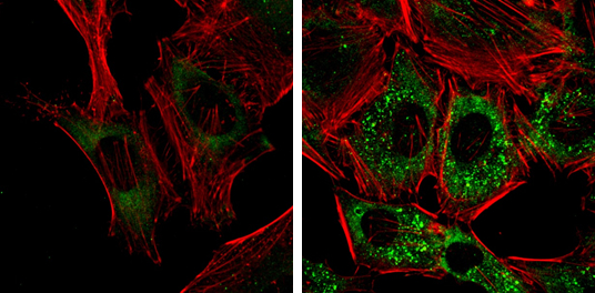
NDP52 antibody [GT1813] detects NDP52 protein at autophagosome by immunofluorescent analysis. Samples: HeLa cells mock (left) and treated with 50μM Chloroquine for 24 hr (right) were fixed in 4% paraformaldehyde at RT for 15 min. Green: NDP52 protein stained by NDP52 antibody [GT1813] (GTX630397) diluted at 1:1000. Red: Phalloidin, a F-actin marker.
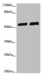
![Non-transfected (–) and transfected (+) HepG2 whole cell extracts (30 μg) were separated by 10% SDS-PAGE, and the membrane was blotted with NDP52 antibody [GT1813] (GTX630397) diluted at 1:1000.](https://www.genetex.com/upload/website/prouct_img/normal/GTX630397/GTX630397_41589_20160721_WB_shRNA_watermark_w_23061202_884.webp "Non-transfected (–) and transfected (+) HepG2 whole cell extracts (30 μg) were separated by 10% SDS-PAGE, and the membrane was blotted with NDP52 antibody [GT1813] (GTX630397) diluted at 1:1000.")
 and treated (+, Thapsigargin treatment for 12hrs and 24hrs) Huh-7 whole cell extracts (30 μg) were separated by 10% SDS-PAGE, and the membrane was blotted with NDP52 antibody (GTX630397) diluted by 1:500. The ACTB was used as internal control (GTX110564, 1:50000) shown at the bottom panel.")
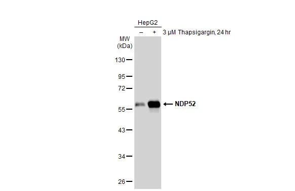
 and treated (+, Thapsigargin treatment for 12hrs and 24hrs) HepG2 whole cell extracts (30 μg) were separated by 10% SDS-PAGE, and the membrane was blotted with NDP52 antibody (GTX630397) diluted by 1:500. The ACTB was used as internal control (GTX110564, 1:50000) shown at the bottom panel.")
![NDP52 antibody [GT1813] detects NDP52 protein by western blot analysis. A. 30 μg Jurkat whole cell lysate/extract B. 30 μg Raji whole cell lysate/extract C. 30 μg NCI-H929 whole cell lysate/extract 10 % SDS-PAGE NDP52 antibody [GT1813] (GTX630397) dilution: 1:1000](https://www.genetex.com/upload/website/prouct_img/normal/GTX630397/GTX630397_41589_WB_w_23061202_730.webp "NDP52 antibody [GT1813] detects NDP52 protein by western blot analysis. A. 30 μg Jurkat whole cell lysate/extract B. 30 μg Raji whole cell lysate/extract C. 30 μg NCI-H929 whole cell lysate/extract 10 % SDS-PAGE NDP52 antibody [GT1813] (GTX630397) dilution: 1:1000")
![NDP52 antibody [GT1813] detects NDP52 protein by western blot analysis. A. 30 μg Huh7 whole cell lysate/extract B. 30 μg Hep3B whole cell lysate/extract C. 30 μg HepG2 whole cell lysate/extract 10 % SDS-PAGE NDP52 antibody [GT1813] (GTX630397) dilution: 1:1000](https://www.genetex.com/upload/website/prouct_img/normal/GTX630397/GTX630397_41589_WB_2_w_23061202_441.webp "NDP52 antibody [GT1813] detects NDP52 protein by western blot analysis. A. 30 μg Huh7 whole cell lysate/extract B. 30 μg Hep3B whole cell lysate/extract C. 30 μg HepG2 whole cell lysate/extract 10 % SDS-PAGE NDP52 antibody [GT1813] (GTX630397) dilution: 1:1000")
NDP52 antibody [GT1813] detects NDP52 protein at autophagosome by immunofluorescent analysis. Samples: HeLa cells mock (left) and treated with 50μM Chloroquine for 24 hr (right) were fixed in 4% paraformaldehyde at RT for 15 min. Green: NDP52 protein stained by NDP52 antibody [GT1813] (GTX630397) diluted at 1:1000. Red: Phalloidin, a F-actin marker.
NDP52 antibody [GT1813]
GTX630397
ApplicationsImmunoFluorescence, Western Blot, ImmunoCytoChemistry
Product group Antibodies
ReactivityHuman
TargetCALCOCO2
Overview
- SupplierGeneTex
- Product NameNDP52 antibody [GT1813]
- Delivery Days Customer9
- Application Supplier NoteWB: 1:500-1:3000. ICC/IF: 1:100-1:1000. *Optimal dilutions/concentrations should be determined by the researcher.Not tested in other applications.
- ApplicationsImmunoFluorescence, Western Blot, ImmunoCytoChemistry
- CertificationResearch Use Only
- ClonalityMonoclonal
- Clone IDGT1813
- Concentration1 mg/ml
- ConjugateUnconjugated
- Gene ID10241
- Target nameCALCOCO2
- Target descriptioncalcium binding and coiled-coil domain 2
- Target synonymsNDP52, calcium-binding and coiled-coil domain-containing protein 2, antigen nuclear dot 52 kDa protein, nuclear domain 10 protein 52, nuclear domain 10 protein NDP52, nuclear dot protein 52
- HostMouse
- IsotypeIgG2a
- Protein IDQ13137
- Protein NameCalcium-binding and coiled-coil domain-containing protein 2
- Scientific DescriptionThe protein encoded by this gene is a subunit of nuclear domain 10 (ND10) bodies. ND10 bodies are nuclear domains appearing immunohistochemically as ten dots per nucleus. They are believed to be associated with the nuclear matrix on the basis of their resistance to nuclease digestion and salt extraction. ND10 proteins are removed from the nucleus by herpes simplex virus-1 infection and may have a role in viral life cycles. [provided by RefSeq]
- ReactivityHuman
- Storage Instruction-20°C or -80°C,2°C to 8°C
- UNSPSC41116161
Datasheet
Related products

Product group Antibodies
Anti-NDP52/CALCOCO2 Antibody Picoband(r)A05876-1-CARRIER-FREE
ApplicationsFlow Cytometry, ImmunoFluorescence, Western Blot, ELISA, ImmunoCytoChemistry, ImmunoHistoChemistry
ReactivityHuman
TargetCALCOCO2
- SizePrice
Product group Antibodies
Anti-CALCOCO2 Antibody144-07358
ApplicationsWestern Blot
ReactivityHuman, Mouse, Rat
TargetCALCOCO2
- SizePrice
Product group Antibodies
CALCOCO2 AntibodyCSB-PA614394ESR1HU
ApplicationsImmunoPrecipitation, Western Blot, ELISA, ImmunoHistoChemistry
ReactivityHuman
TargetCALCOCO2
- SizePrice
![ICC/IF analysis of PFA-fixed U2OS cells using GTX04863 NDP52 antibody [9E2F2]. Orange : Primary antibody Blue : DAPI Antigen retrieval : IHC enzyme antigen retrieval reagent for 15 mins Dilution : 5 μg/mL](https://www.genetex.com/upload/website/prouct_img/normal/GTX04863/GTX04863_20240531_ICCIF_24053100_480.webp)
Product group Antibodies
NDP52 antibody [9E2F2]GTX04863
ApplicationsFlow Cytometry, ImmunoFluorescence, Western Blot, ImmunoCytoChemistry
ReactivityHuman, Mouse, Rat
TargetCALCOCO2
- SizePrice
Product group Antibodies

CALCOCO2 AntibodyLS-C349083
ApplicationsWestern Blot, ImmunoHistoChemistry
ReactivityHuman, Mouse, Rat
TargetCALCOCO2
- SizePrice

Product group Antibodies
Anti-CALCOCO2 AntibodyHPA023195
ApplicationsWestern Blot, ImmunoCytoChemistry, ImmunoHistoChemistry
ReactivityHuman
TargetCALCOCO2
- SizePrice
Product group Antibodies
NDP52 antibodyGTX115378
ApplicationsImmunoFluorescence, ImmunoPrecipitation, Western Blot, ImmunoCytoChemistry, ImmunoHistoChemistry, ImmunoHistoChemistry Paraffin
ReactivityHuman
TargetCALCOCO2
- SizePrice
![NDP52 antibody [HL3126] detects NDP52 protein by immunohistochemical analysis. Sample: Paraffin-embedded human breast carcinoma. NDP52 stained by NDP52 antibody [HL3126] (GTX640612) diluted at 1:100. Antigen Retrieval: Citrate buffer, pH 6.0, 15 min](https://www.genetex.com/upload/website/prouct_img/normal/GTX640612/GTX640612_T-45467_20240806_IHC-P_24082802_845.webp)
Product group Antibodies
NDP52 antibody [HL3126]GTX640612
ApplicationsWestern Blot, ImmunoHistoChemistry, ImmunoHistoChemistry Paraffin
ReactivityHuman
TargetCALCOCO2
- SizePrice
Product group Antibodies
NDP52 antibodyGTX55720
ApplicationsImmunoFluorescence, Western Blot, ImmunoCytoChemistry
ReactivityHuman, Mouse, Rat
TargetCALCOCO2
- SizePrice

Product group Antibodies
NDP52 antibody [GT422]GTX630396
ApplicationsImmunoFluorescence, ImmunoPrecipitation, Western Blot, ImmunoCytoChemistry
ReactivityHuman
TargetCALCOCO2
- SizePrice