

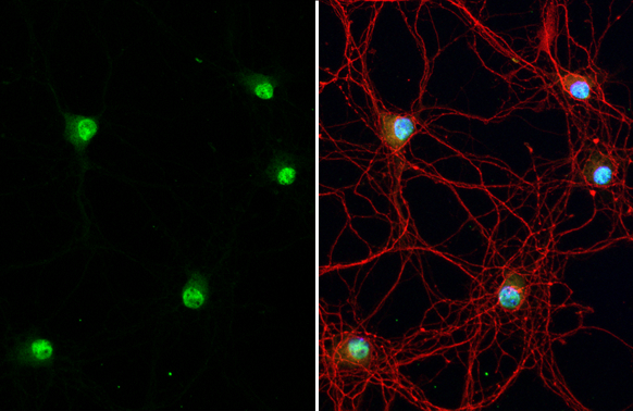
NeuN antibody detects NeuN protein at nucleus by immunofluorescent analysis. Sample: DIV9 rat E18 primary hippocampal neuron cells were fixed in 4% paraformaldehyde at RT for 15 min. Green: NeuN stained by NeuN antibody (GTX132974) diluted at 1:250. Red: Tau, an axon marker, stained by Tau antibody [GT287] (GTX634809) diluted at 1:500. Blue: Fluoroshield with DAPI (GTX30920).
![NeuN antibody detects NeuN protein at nucleus by immunofluorescent analysis. Sample: DIV9 rat E18 primary cortical neurons were fixed in 4% paraformaldehyde at RT for 15 min. Green: NeuN protein stained by NeuN antibody (GTX132974) diluted at 1:1000. Red: beta Tubulin 3/ Tuj1, stained by beta Tubulin 3/ Tuj1 antibody [GT886] (GTX631830) diluted at 1:500. Blue: Fluoroshield with DAPI (GTX30920).](https://www.genetex.com/upload/website/prouct_img/normal/GTX132974/GTX132974_42564_20170503_IFA_R_w_23060523_597.webp "NeuN antibody detects NeuN protein at nucleus by immunofluorescent analysis. Sample: DIV9 rat E18 primary cortical neurons were fixed in 4% paraformaldehyde at RT for 15 min. Green: NeuN protein stained by NeuN antibody (GTX132974) diluted at 1:1000. Red: beta Tubulin 3/ Tuj1, stained by beta Tubulin 3/ Tuj1 antibody [GT886] (GTX631830) diluted at 1:500. Blue: Fluoroshield with DAPI (GTX30920).")
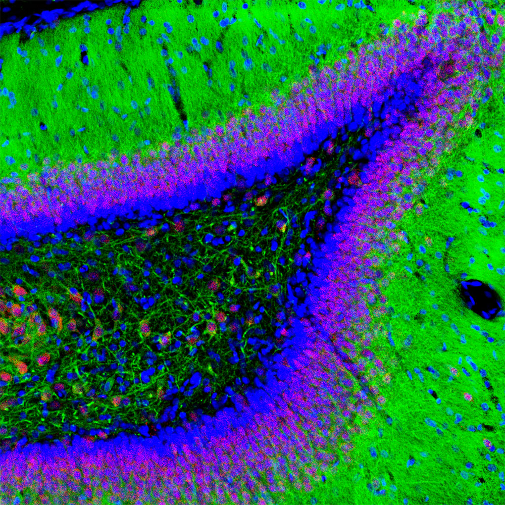
![NeuN antibody detects NeuN protein at nucleus by immunohistochemical analysis. Sample: Paraffin-embedded 3xTg-AD transgenic mouse brain (left) and healthy mouse brain (right). Red: NeuN stained by NeuN antibody (GTX132974) diluted at 1:250. Green: Beta amyloid (1-42) stained by Beta amyloid (1-42) antibody – Conformation Specific antibody [GT622] (GTX635160) diluted at 1:1000. Blue: Fluoroshield with DAPI (GTX30920). Antigen Retrieval: Citrate buffer, pH 6.0, 15 min This image was provided courtesy of a customer review.](https://www.genetex.com/upload/website/prouct_img/normal/GTX132974/GTX132974_42564_20190510_IHC-P-FL_M_w_23060523_297.webp "NeuN antibody detects NeuN protein at nucleus by immunohistochemical analysis. Sample: Paraffin-embedded 3xTg-AD transgenic mouse brain (left) and healthy mouse brain (right). Red: NeuN stained by NeuN antibody (GTX132974) diluted at 1:250. Green: Beta amyloid (1-42) stained by Beta amyloid (1-42) antibody – Conformation Specific antibody [GT622] (GTX635160) diluted at 1:1000. Blue: Fluoroshield with DAPI (GTX30920). Antigen Retrieval: Citrate buffer, pH 6.0, 15 min This image was provided courtesy of a customer review.")
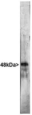
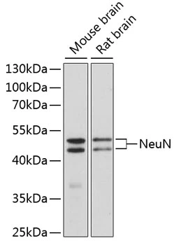
 were separated by 10% SDS-PAGE, and the membrane was blotted with NeuN antibody (GTX132974) diluted at 1:1000. The HRP-conjugated anti-rabbit IgG antibody (GTX213110-01) was used to detect the primary antibody.")
![NeuN antibody detects NeuN protein expression by immunohistochemical analysis. Sample: Frozen-sectioned adult mouse cerebellum. Green: NeuN protein stained by NeuN antibody (GTX132974) diluted at 1:250. Red: beta Tubulin 3/ TUJ1, stained by beta Tubulin 3/ TUJ1 antibody [GT11710] (GTX631836) diluted at 1:500. Blue: Fluoroshield with DAPI (GTX30920).](https://www.genetex.com/upload/website/prouct_img/normal/GTX132974/GTX132974_42564_20170531_IHC-Fr_M_w_23060523_819.webp "NeuN antibody detects NeuN protein expression by immunohistochemical analysis. Sample: Frozen-sectioned adult mouse cerebellum. Green: NeuN protein stained by NeuN antibody (GTX132974) diluted at 1:250. Red: beta Tubulin 3/ TUJ1, stained by beta Tubulin 3/ TUJ1 antibody [GT11710] (GTX631836) diluted at 1:500. Blue: Fluoroshield with DAPI (GTX30920).")
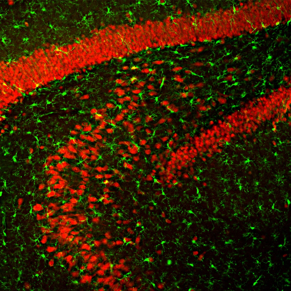
![NeuN antibody detects NeuN protein expression by immunohistochemical analysis. Sample: Frozen-sectioned adult mouse hippocampus. Green: NeuN protein stained by NeuN antibody (GTX132974) diluted at 1:250. Red: alpha Tubulin, stained by alpha Tubulin antibody [GT114] (GTX628802) diluted at 1:500.](https://www.genetex.com/upload/website/prouct_img/normal/GTX132974/GTX132974_42564_42886_IHC-Fr_M_w_23060523_317.webp "NeuN antibody detects NeuN protein expression by immunohistochemical analysis. Sample: Frozen-sectioned adult mouse hippocampus. Green: NeuN protein stained by NeuN antibody (GTX132974) diluted at 1:250. Red: alpha Tubulin, stained by alpha Tubulin antibody [GT114] (GTX628802) diluted at 1:500.")
 diluted at 1:500. Antigen Retrieval: Citrate buffer, pH 6.0, 15 min")
 diluted at 1:500. Antigen Retrieval: Citrate buffer, pH 6.0, 15 min")
 diluted at 1:500. Antigen Retrieval: Citrate buffer, pH 6.0, 15 min")
 diluted at 1:500. Antigen Retrieval: Citrate buffer, pH 6.0, 15 min")
NeuN antibody detects NeuN protein at nucleus by immunofluorescent analysis. Sample: DIV9 rat E18 primary hippocampal neuron cells were fixed in 4% paraformaldehyde at RT for 15 min. Green: NeuN stained by NeuN antibody (GTX132974) diluted at 1:250. Red: Tau, an axon marker, stained by Tau antibody [GT287] (GTX634809) diluted at 1:500. Blue: Fluoroshield with DAPI (GTX30920).
NeuN antibody
GTX132974
ApplicationsImmunoFluorescence, Western Blot, ImmunoCytoChemistry, ImmunoHistoChemistry, ImmunoHistoChemistry Frozen, ImmunoHistoChemistry Paraffin
Product group Antibodies
ReactivityFish, Human, Mouse, Rat
TargetRBFOX3
Overview
- SupplierGeneTex
- Product NameNeuN antibody
- Delivery Days Customer9
- Application Supplier NoteWB: 1:500-1:3000. ICC/IF: 1:100-1:1000. IHC-P: 1:100-1:1000. IHC-Fr: 1:100-1:1000. *Optimal dilutions/concentrations should be determined by the researcher.Not tested in other applications.
- ApplicationsImmunoFluorescence, Western Blot, ImmunoCytoChemistry, ImmunoHistoChemistry, ImmunoHistoChemistry Frozen, ImmunoHistoChemistry Paraffin
- CertificationResearch Use Only
- ClonalityPolyclonal
- Concentration1.32 mg/ml
- ConjugateUnconjugated
- Gene ID146713
- Target nameRBFOX3
- Target descriptionRNA binding fox-1 homolog 3
- Target synonymsFOX-3, FOX3, HRNBP3, NEUN, RNA binding protein fox-1 homolog 3, RNA binding protein, fox-1 homolog 3, fox-1 homolog C, hexaribonucleotide binding protein 3, neuN antigen, neuronal nuclei antigen
- HostRabbit
- IsotypeIgG
- Protein IDA6NFN3
- Protein NameRNA binding protein fox-1 homolog 3
- ReactivityFish, Human, Mouse, Rat
- Storage Instruction-20°C or -80°C,2°C to 8°C
- UNSPSC41116161
Datasheet
Related products
Product group Antibodies
Anti-NeuN AntibodyA270544
ApplicationsImmunoFluorescence, Western Blot, ImmunoCytoChemistry, ImmunoHistoChemistry
ReactivityHuman, Mouse, Rat
- SizePrice

Product group Antibodies
Anti-RBFOX3 AntibodyAMAB91746
ApplicationsImmunoHistoChemistry
ReactivityHuman, Mouse
TargetRBFOX3
- SizePrice

Product group Antibodies
References
ApplicationsFlow Cytometry, ImmunoFluorescence, Western Blot, ImmunoCytoChemistry, ImmunoHistoChemistry
ReactivityHuman, Mouse, Rat
TargetRBFOX3
- SizePrice
Product group Antibodies
References
NeuN Polyclonal AntibodyBS-1613R
ApplicationsFlow Cytometry, ImmunoFluorescence, Western Blot, ELISA, ImmunoCytoChemistry, ImmunoHistoChemistry, ImmunoHistoChemistry Frozen, ImmunoHistoChemistry Paraffin
ReactivityBovine, Canine, Equine, Human, Mouse, Rat
TargetRBFOX3
- SizePrice
Product group Antibodies
RBFOX3 AntibodyCSB-PA070474
ApplicationsWestern Blot, ELISA, ImmunoHistoChemistry
ReactivityHuman, Mouse
TargetRBFOX3
- SizePrice
Product group Antibodies
References
NeuN antibodyGTX16208
ApplicationsImmunoFluorescence, Western Blot, ImmunoCytoChemistry, ImmunoHistoChemistry, ImmunoHistoChemistry Frozen
ReactivityBovine, Chicken, Equine, Human, Mouse, Porcine, Rat
TargetRBFOX3
- SizePrice
Product group Antibodies
References
NeuN antibodyGTX00837
ApplicationsImmunoFluorescence, Western Blot, ImmunoCytoChemistry, ImmunoHistoChemistry, ImmunoHistoChemistry Frozen, ImmunoHistoChemistry Paraffin
ReactivityHuman, Mouse, Rat
TargetRBFOX3
- SizePrice
Product group Antibodies

RBFOX3 / NEUN AntibodyLS-C335111
ApplicationsImmunoFluorescence, Western Blot, ImmunoHistoChemistry
ReactivityHuman, Mouse, Rat
TargetRBFOX3
- SizePrice
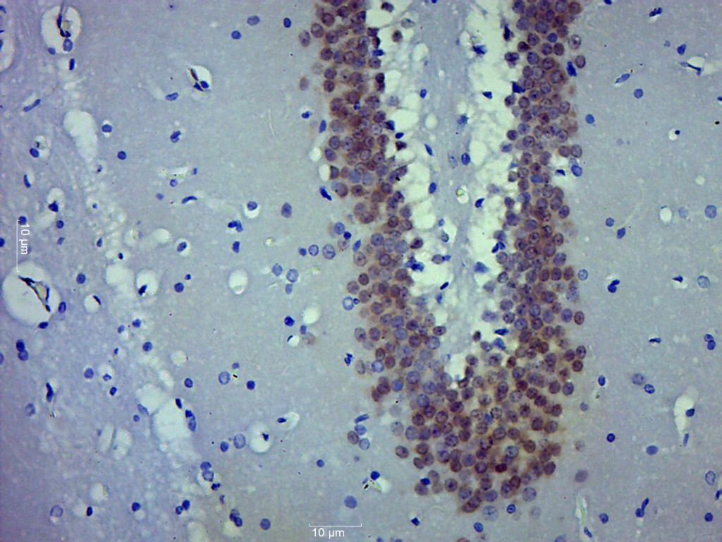
![NeuN antibody [HL2194] detects NeuN protein by immunohistochemical analysis. Sample: Paraffin-embedded rat tissues. NeuN stained by NeuN antibody [HL2194] (GTX638198) diluted at 1:100. Antigen Retrieval: Citrate buffer, pH 6.0, 15 min](https://www.genetex.com/upload/website/prouct_img/normal/GTX638198/GTX638198_T-44942_20230424_IHC-P_multiple_R_23050918_845.webp)
Product group Antibodies
NeuN antibody [HL2194]GTX638198
ApplicationsImmunoFluorescence, ImmunoCytoChemistry, ImmunoHistoChemistry, ImmunoHistoChemistry Frozen, ImmunoHistoChemistry Paraffin
ReactivityHuman, Mouse, Rat
TargetRBFOX3
- SizePrice
Product group Antibodies
NeuN antibodyGTX32746
ApplicationsWestern Blot, ImmunoHistoChemistry, ImmunoHistoChemistry Paraffin
ReactivityHuman, Mouse, Rat
TargetRBFOX3
- SizePrice