

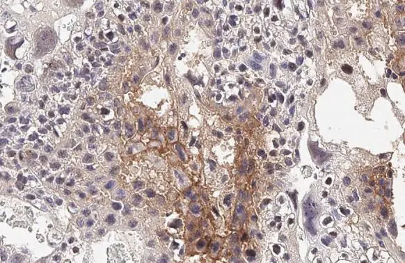
PD-L1 antibody [HL2293] detects PD-L1 protein at cell membrane and nucleus by immunohistochemical analysis. Sample: Paraffin-embedded mouse placenta. PD-L1 stained by PD-L1 antibody [HL2293] (GTX638348) diluted at 1:1000. Antigen Retrieval: Citrate buffer, pH 6.0, 15 min
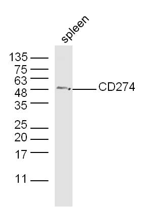
![Mouse tissue extract (50 μg) was separated by 10% SDS-PAGE, and the membrane was blotted with PD-L1 antibody [HL2293] (GTX638348) diluted at 1:2000. The HRP-conjugated anti-rabbit IgG antibody (GTX213110-01) was used to detect the primary antibody.](https://www.genetex.com/upload/website/prouct_img/normal/GTX638348/GTX638348_45145_20230908_WB_M_thymus_23091319_904.webp "Mouse tissue extract (50 μg) was separated by 10% SDS-PAGE, and the membrane was blotted with PD-L1 antibody [HL2293] (GTX638348) diluted at 1:2000. The HRP-conjugated anti-rabbit IgG antibody (GTX213110-01) was used to detect the primary antibody.")
PD-L1 antibody [HL2293] detects PD-L1 protein at cell membrane and nucleus by immunohistochemical analysis. Sample: Paraffin-embedded mouse placenta. PD-L1 stained by PD-L1 antibody [HL2293] (GTX638348) diluted at 1:1000. Antigen Retrieval: Citrate buffer, pH 6.0, 15 min
PD-L1 antibody [HL2293]
GTX638348
ApplicationsWestern Blot, ImmunoHistoChemistry, ImmunoHistoChemistry Paraffin
Product group Antibodies
ReactivityMouse
TargetCd274
Overview
- SupplierGeneTex
- Product NamePD-L1 antibody [HL2293]
- Delivery Days Customer9
- Application Supplier NoteWB: 1:500-1:3000. IHC-P: 1:100-1:1000. *Optimal dilutions/concentrations should be determined by the researcher.Not tested in other applications.
- ApplicationsWestern Blot, ImmunoHistoChemistry, ImmunoHistoChemistry Paraffin
- CertificationResearch Use Only
- ClonalityMonoclonal
- Clone IDHL2293
- Concentration1 mg/ml
- ConjugateUnconjugated
- Gene ID60533
- Target nameCd274
- Target descriptionCD274 antigen
- Target synonymsA530045L16Rik, B7h1, Pdcd1l1, Pdcd1lg1, Pdl1, programmed cell death 1 ligand 1, B7 homolog 1, PDCD1 ligand 1
- HostRabbit
- IsotypeIgG
- Protein IDQ9EP73
- Protein NameProgrammed cell death 1 ligand 1
- Scientific DescriptionThe protein encoded by this gene is an immune inhibitory receptor ligand that is expressed by hematopoietic and non-hematopoietic cells, such as T cells and B cells and various types of tumor cells. The encoded protein is a type I transmembrane protein that has immunoglobulin V-like and C-like domains. Interaction of this ligand with its receptor inhibits T-cell activation and cytokine production. During infection or inflammation of normal tissue, this interaction is important for preventing autoimmunity by maintaining homeostasis of the immune response. In tumor microenvironments, this interaction provides an immune escape for tumor cells through cytotoxic T-cell inactivation. Mice deficient for this gene display a variety of phenotypes including decreased allogeneic fetal survival rates and severe experimental autoimmune encephalomyelitis. [provided by RefSeq, Sep 2015]
- ReactivityMouse
- Storage Instruction-20°C or -80°C,2°C to 8°C
- UNSPSC41116161
Datasheet
Related products

Product group Antibodies
Anti-PD-L1/Cd274 Antibody Picoband(r)A00109-3-CARRIER-FREE
ApplicationsWestern Blot, ELISA
ReactivityMouse
TargetCd274
- SizePrice
Product group Antibodies
ApplicationsFlow Cytometry
ReactivityMouse
TargetCd274
- SizePrice
Product group Antibodies

Anti-PD-L1 [10F.9G2]AB01419-3.0-VXS
ApplicationsFlow Cytometry, ELISA, ImmunoHistoChemistry, Neutralisation/Blocking
ReactivityMouse
TargetCd274
- SizePrice
Product group Antibodies
PD-L1 antibody [MAB0871]GTX52447
ApplicationsFlow Cytometry, Neutralisation/Blocking
ReactivityMouse
TargetCd274
- SizePrice
Product group Antibodies
PD-L1 Polyclonal AntibodyBS-1103R
ApplicationsImmunoFluorescence, Western Blot, ELISA, ImmunoCytoChemistry, ImmunoHistoChemistry, ImmunoHistoChemistry Frozen, ImmunoHistoChemistry Paraffin
ReactivityBovine, Equine, Human, Mouse, Porcine, Rat, Sheep
TargetCd274
- SizePrice
![WB analysis of purified PD-L1 recombinant protein using GTX14145 PD-L1 antibody [10F.9G2]. Lane 1 : 0.1 ug reduced purified mouse PD-L1 with histidine tag at C-terminuse Lane 2 : 0.05 ug reduced purified mousePD-L1 with histidine tag at C-terminus Dilution : 8 ug/ml](https://www.genetex.com/upload/website/prouct_img/normal/GTX14145/GTX14145_20200917_WB_w_23060620_268.webp)
Product group Antibodies
PD-L1 antibody [10F.9G2]GTX14145
ApplicationsFlow Cytometry, ImmunoFluorescence, Western Blot, ImmunoCytoChemistry, ImmunoHistoChemistry, ImmunoHistoChemistry Frozen, Neutralisation/Blocking
ReactivityMouse
TargetCd274
- SizePrice
Product group Antibodies
anti-PD-L1 (human), Rabbit Monoclonal (RM320)REV-31-1205-00
ApplicationsWestern Blot, ImmunoHistoChemistry
ReactivityHuman
TargetCd274
- SizePrice
Product group Antibodies
ApplicationsImmunoPrecipitation, Western Blot, ImmunoCytoChemistry, ImmunoHistoChemistry
ReactivityMouse
TargetCd274
- SizePrice