

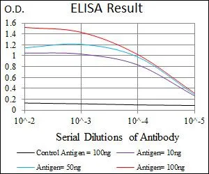
ELISA analysis of antigen using GTX60627 PLK1 antibody [3C11]. Black : Control antigen 100ng Purple : Antigen 10ng Blue : Antigen 50ng Red : Antigen 100ng
![FACS analysis of NIH3T3 cells using GTX60627 PLK1 antibody [3C11]. Green : PLK1 Red : negative control](https://www.genetex.com/upload/website/prouct_img/normal/GTX60627/GTX60627_20170912_FACS_w_23061123_117.webp "FACS analysis of NIH3T3 cells using GTX60627 PLK1 antibody [3C11]. Green : PLK1 Red : negative control")
![IHC-P analysis of stomach cancer tissue using GTX60627 PLK1 antibody [3C11].](https://www.genetex.com/upload/website/prouct_img/normal/GTX60627/GTX60627_20170912_IHC-P_1_w_23061123_799.webp "IHC-P analysis of stomach cancer tissue using GTX60627 PLK1 antibody [3C11].")
![IHC-P analysis of rectum cancer tissue using GTX60627 PLK1 antibody [3C11].](https://www.genetex.com/upload/website/prouct_img/normal/GTX60627/GTX60627_20170912_IHC-P_w_23061123_657.webp "IHC-P analysis of rectum cancer tissue using GTX60627 PLK1 antibody [3C11].")
![WB analysis of K562 (1) and Raji (2) cell lysate using GTX60627 PLK1 antibody [3C11].](https://www.genetex.com/upload/website/prouct_img/normal/GTX60627/GTX60627_20170912_WB_w_23061123_843.webp "WB analysis of K562 (1) and Raji (2) cell lysate using GTX60627 PLK1 antibody [3C11].")
ELISA analysis of antigen using GTX60627 PLK1 antibody [3C11]. Black : Control antigen 100ng Purple : Antigen 10ng Blue : Antigen 50ng Red : Antigen 100ng
PLK1 antibody [3C11]
GTX60627
ApplicationsFlow Cytometry, Western Blot, ELISA, ImmunoHistoChemistry, ImmunoHistoChemistry Paraffin
Product group Antibodies
ReactivityHuman, Mouse
TargetPLK1
Overview
- SupplierGeneTex
- Product NamePLK1 antibody [3C11]
- Delivery Days Customer9
- Application Supplier NoteWB: 1/500 - 1/2000. IHC-P: 1/200 - 1/1000. FACS: 1/200 - 1/400. ELISA: 1/10000. *Optimal dilutions/concentrations should be determined by the researcher.Not tested in other applications.
- ApplicationsFlow Cytometry, Western Blot, ELISA, ImmunoHistoChemistry, ImmunoHistoChemistry Paraffin
- CertificationResearch Use Only
- ClonalityMonoclonal
- Clone ID3C11
- Concentration1 mg/ml
- ConjugateUnconjugated
- Gene ID5347
- Target namePLK1
- Target descriptionpolo like kinase 1
- Target synonymsPLK, STPK13, serine/threonine-protein kinase PLK1, PLK-1, cell cycle regulated protein kinase, polo (Drosophia)-like kinase, serine/threonine-protein kinase 13
- HostMouse
- IsotypeIgG1
- Protein IDP53350
- Protein NameSerine/threonine-protein kinase PLK1
- ReactivityHuman, Mouse
- Storage Instruction-20°C or -80°C,2°C to 8°C
- UNSPSC12352203
Datasheet
Related products

Product group Antibodies
Anti-PLK1 Antibody Picoband(r)PB9500-CARRIER-FREE
ApplicationsWestern Blot
ReactivityHuman
TargetPLK1
- SizePrice
Product group Antibodies
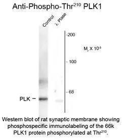
PLK1 (phospho Thr210) antibodyGTX82612
ApplicationsWestern Blot
ReactivityHuman, Rat
TargetPLK1
- SizePrice
![FACS analysis of HEK293T cells transfected with either PLK1 plasmid(Red) or empty vector control plasmid(Blue) using GTX83864 PLK1 antibody [3F12].](https://www.genetex.com/upload/website/prouct_img/normal/GTX83864/GTX83864_204_FACS_w_23061420_168.webp)
Product group Antibodies
PLK1 antibody [3F12]GTX83864
ApplicationsFlow Cytometry, ImmunoPrecipitation, Western Blot
ReactivityHuman, Mouse
TargetPLK1
- SizePrice
![IHC-P analysis of human breast adenocarcinoma tissue using GTX83865 PLK1 antibody [1D4]. Antigen retrieval : Heat-induced epitope retrieval by 10mM citrate buffer, pH6.0, 100oC for 10min.](https://www.genetex.com/upload/website/prouct_img/normal/GTX83865/GTX83865_1943_IHC-P_w_23061420_628.webp)
Product group Antibodies
PLK1 antibody [1D4]GTX83865
ApplicationsFlow Cytometry, ImmunoPrecipitation, Western Blot, ImmunoHistoChemistry, ImmunoHistoChemistry Paraffin
ReactivityHuman
TargetPLK1
- SizePrice
![ICC/IF analysis of COS7 cells transiently transfected with PLK1 plasmid using GTX83866 PLK1 antibody [8C12]. Dilution : 1:100](https://www.genetex.com/upload/website/prouct_img/normal/GTX83866/GTX83866_1230_ICCIF_w_23061420_550.webp)
Product group Antibodies
PLK1 antibody [8C12]GTX83866
ApplicationsFlow Cytometry, ImmunoFluorescence, ImmunoPrecipitation, Western Blot, ImmunoCytoChemistry
ReactivityCanine, Human, Monkey
TargetPLK1
- SizePrice
Product group Antibodies
Anti-Phospho-PLK1-T210 Antibody144-50717
ApplicationsWestern Blot
ReactivityHuman
TargetPLK1
- SizePrice
![ELISA analysis of antigen using GTX60624 PLK1 antibody [1D1]. Black : Control antigen 100ng Purple : Antigen 10ng Blue : Antigen 50ng Red : Antigen 100ng](https://www.genetex.com/upload/website/prouct_img/normal/GTX60624/GTX60624_20170912_ELISA_w_23061123_595.webp)
Product group Antibodies
PLK1 antibody [1D1]GTX60624
ApplicationsImmunoFluorescence, Western Blot, ELISA, ImmunoCytoChemistry
ReactivityHuman
TargetPLK1
- SizePrice
Product group Antibodies

Goat anti-PLK1 AntibodyEB06969
ApplicationsWestern Blot, ELISA
ReactivityCanine, Human, Mouse, Rat
TargetPLK1
- SizePrice
![ICC/IF analysis of HeLa cells using GTX14210 PLK1 antibody [Mixed clones]. Dilution : 1microg/ml](https://www.genetex.com/upload/website/prouct_img/normal/GTX14210/GTX14210_20191203_ICC-IF_3_w_23060620_538.webp)
Product group Antibodies
PLK1 antibody [Mixed clones]GTX14210
ApplicationsFlow Cytometry, ImmunoFluorescence, ImmunoPrecipitation, Western Blot, ELISA, ImmunoCytoChemistry, ImmunoHistoChemistry, ImmunoHistoChemistry Paraffin
ReactivityHuman, Monkey, Mouse, Rat, Xenopus
TargetPLK1
- SizePrice
Product group Antibodies
PLK1 (phospho Thr210) antibodyGTX03368
ApplicationsImmunoFluorescence, Western Blot, ELISA, ImmunoCytoChemistry, ImmunoHistoChemistry, ImmunoHistoChemistry Paraffin
ReactivityHuman, Mouse
TargetPLK1
- SizePrice