

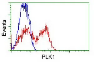
FACS analysis of HEK293T cells transfected with either PLK1 plasmid(Red) or empty vector control plasmid(Blue) using GTX83864 PLK1 antibody [3F12].
![IP analysis of DDDDK tagged PLK1 overexpressed HEK293T lysate using GTX83864 PLK1 antibody [3F12].After extensive wash to remove any non-specific binding, the immuno-precipitated products were analyzed with rabbit anti-DDDDK polyclonal antibody. IP reaction : 2microg antibody / 500microl cell lysate](https://www.genetex.com/upload/website/prouct_img/normal/GTX83864/GTX83864_3385_IP_w_23061420_386.webp "IP analysis of DDDDK tagged PLK1 overexpressed HEK293T lysate using GTX83864 PLK1 antibody [3F12].After extensive wash to remove any non-specific binding, the immuno-precipitated products were analyzed with rabbit anti-DDDDK polyclonal antibody. IP reaction : 2microg antibody / 500microl cell lysate")
![WB analysis of various cell lines using GTX83864 PLK1 antibody [3F12]. Loading : 35 ug per lane Dilution : 1:200](https://www.genetex.com/upload/website/prouct_img/normal/GTX83864/GTX83864_3464_WB_w_23061420_283.webp "WB analysis of various cell lines using GTX83864 PLK1 antibody [3F12]. Loading : 35 ug per lane Dilution : 1:200")
![WB analysis of HEK293T cells transfected with PLK1 plasmid (Right) or empty vector (Left) for 48 hrs using GTX83864 PLK1 antibody [3F12]. Loading : 5 ug per lane Dilution : 1:500](https://www.genetex.com/upload/website/prouct_img/normal/GTX83864/GTX83864_4395_WB_w_23061420_379.webp "WB analysis of HEK293T cells transfected with PLK1 plasmid (Right) or empty vector (Left) for 48 hrs using GTX83864 PLK1 antibody [3F12]. Loading : 5 ug per lane Dilution : 1:500")
FACS analysis of HEK293T cells transfected with either PLK1 plasmid(Red) or empty vector control plasmid(Blue) using GTX83864 PLK1 antibody [3F12].
PLK1 antibody [3F12]
GTX83864
ApplicationsFlow Cytometry, ImmunoPrecipitation, Western Blot
Product group Antibodies
ReactivityHuman, Mouse
TargetPLK1
Overview
- SupplierGeneTex
- Product NamePLK1 antibody [3F12]
- Delivery Days Customer9
- Application Supplier NoteWB: 1:1000-1:2000. FACS: 1:100. IP: 2ug/500ul. *Optimal dilutions/concentrations should be determined by the researcher.Not tested in other applications.
- ApplicationsFlow Cytometry, ImmunoPrecipitation, Western Blot
- CertificationResearch Use Only
- ClonalityMonoclonal
- Clone ID3F12
- Concentration1.01 mg/ml
- ConjugateUnconjugated
- Gene ID5347
- Target namePLK1
- Target descriptionpolo like kinase 1
- Target synonymsPLK, STPK13, serine/threonine-protein kinase PLK1, PLK-1, cell cycle regulated protein kinase, polo (Drosophia)-like kinase, serine/threonine-protein kinase 13
- HostMouse
- IsotypeIgG1
- Protein IDP53350
- Protein NameSerine/threonine-protein kinase PLK1
- Scientific DescriptionSerine/threonine-protein kinase that performs several important functions throughout M phase of the cell cycle, including the regulation of centrosome maturation and spindle assembly, the removal of cohesins from chromosome arms, the inactivation of APC/C inhibitors, and the regulation of mitotic exit and cytokinesis. Required for recovery after DNA damage checkpoint and entry into mitosis. Required for kinetochore localization of BUB1B. Phosphorylates SGOL1. Required for spindle pole localization of isoform 3 of SGOL1 and plays a role in regulating its centriole cohesion function. Phosphorylates BORA, and thereby promotes the degradation of BORA. Contributes to the regulation of AURKA function. Regulates TP53 stability through phosphorylation of TOPORS.
- ReactivityHuman, Mouse
- Storage Instruction-20°C or -80°C,2°C to 8°C
- UNSPSC12352203
Datasheet
Related products
Product group Antibodies
References
PLK1 Polyclonal AntibodyBS-3535R
ApplicationsImmunoFluorescence, Western Blot, ELISA, ImmunoCytoChemistry, ImmunoHistoChemistry, ImmunoHistoChemistry Frozen, ImmunoHistoChemistry Paraffin
ReactivityCanine, Human, Mouse, Porcine, Rabbit, Rat
TargetPLK1
- SizePrice
Product group Antibodies
Anti-PLK1 AntibodyA85042
ApplicationsWestern Blot, ELISA
ReactivityHuman
- SizePrice
Product group Antibodies
Anti-Phospho-PLK1-T210 Antibody144-50717
ApplicationsWestern Blot
ReactivityHuman
TargetPLK1
- SizePrice

Product group Antibodies
Anti-PLK1 AntibodyAMAB91515
ApplicationsWestern Blot
ReactivityHuman
TargetPLK1
- SizePrice
Product group Antibodies

Goat anti-PLK1 AntibodyEB06969
ApplicationsWestern Blot, ELISA
ReactivityCanine, Human, Mouse, Rat
TargetPLK1
- SizePrice
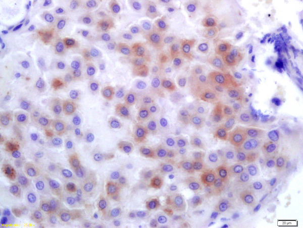
![IHC-P analysis of squamous cell carcinoma of oropharangeal tissue using GTX01927 PLK1 antibody [MJS1]. Note the intense nuclear and cytoplasmic staining of a proportion of proliferating malignant cells.](https://www.genetex.com/upload/website/prouct_img/normal/GTX01927/GTX01927_20200811_IHC-P_73_w_23053121_314.webp)
Product group Antibodies
PLK1 antibody [MJS1]GTX01927
ApplicationsImmunoHistoChemistry, ImmunoHistoChemistry Paraffin
ReactivityHuman
TargetPLK1
- SizePrice
Product group Antibodies
PLK1 (phospho Thr210) antibodyGTX82612
ApplicationsWestern Blot
ReactivityHuman, Rat
TargetPLK1
- SizePrice
![IHC-P analysis of human breast adenocarcinoma tissue using GTX83865 PLK1 antibody [1D4]. Antigen retrieval : Heat-induced epitope retrieval by 10mM citrate buffer, pH6.0, 100oC for 10min.](https://www.genetex.com/upload/website/prouct_img/normal/GTX83865/GTX83865_1943_IHC-P_w_23061420_628.webp)
Product group Antibodies
PLK1 antibody [1D4]GTX83865
ApplicationsFlow Cytometry, ImmunoPrecipitation, Western Blot, ImmunoHistoChemistry, ImmunoHistoChemistry Paraffin
ReactivityHuman
TargetPLK1
- SizePrice
![ICC/IF analysis of COS7 cells transiently transfected with PLK1 plasmid using GTX83866 PLK1 antibody [8C12]. Dilution : 1:100](https://www.genetex.com/upload/website/prouct_img/normal/GTX83866/GTX83866_1230_ICCIF_w_23061420_550.webp)
Product group Antibodies
PLK1 antibody [8C12]GTX83866
ApplicationsFlow Cytometry, ImmunoFluorescence, ImmunoPrecipitation, Western Blot, ImmunoCytoChemistry
ReactivityCanine, Human, Monkey
TargetPLK1
- SizePrice