

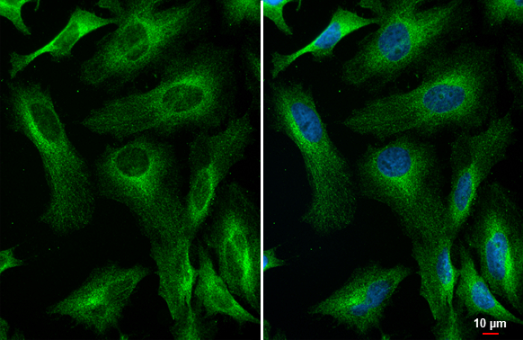
RIP antibody [HL2249] detects RIP protein at endoplasmic reticulum and cytoplasm by immunofluorescent analysis. Sample: HeLa cells were fixed in 4% paraformaldehyde at RT for 15 min. Green: RIP stained by RIP antibody [HL2249] (GTX638298) diluted at 1:500. Blue: Fluoroshield with DAPI (GTX30920).
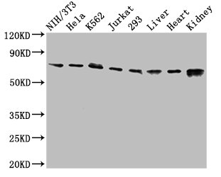
![Various whole cell extracts (30 μg) were separated by 7.5% SDS-PAGE, and the membrane was blotted with RIP antibody [HL2249] (GTX638298) diluted at 1:1000. The HRP-conjugated anti-rabbit IgG antibody (GTX213110-01) was used to detect the primary antibody.](https://www.genetex.com/upload/website/prouct_img/normal/GTX638298/GTX638298_45061_20230602_WB_23060622_474.webp "Various whole cell extracts (30 μg) were separated by 7.5% SDS-PAGE, and the membrane was blotted with RIP antibody [HL2249] (GTX638298) diluted at 1:1000. The HRP-conjugated anti-rabbit IgG antibody (GTX213110-01) was used to detect the primary antibody.")
RIP antibody [HL2249] detects RIP protein at endoplasmic reticulum and cytoplasm by immunofluorescent analysis. Sample: HeLa cells were fixed in 4% paraformaldehyde at RT for 15 min. Green: RIP stained by RIP antibody [HL2249] (GTX638298) diluted at 1:500. Blue: Fluoroshield with DAPI (GTX30920).
RIP antibody [HL2249]
GTX638298
ApplicationsImmunoFluorescence, Western Blot, ImmunoCytoChemistry
Product group Antibodies
ReactivityHuman
TargetRIPK1
Overview
- SupplierGeneTex
- Product NameRIP antibody [HL2249]
- Delivery Days Customer9
- Application Supplier NoteWB: 1:500-1:3000. ICC/IF: 1:100-1:1000. *Optimal dilutions/concentrations should be determined by the researcher.Not tested in other applications.
- ApplicationsImmunoFluorescence, Western Blot, ImmunoCytoChemistry
- CertificationResearch Use Only
- ClonalityMonoclonal
- Clone IDHL2249
- Concentration1 mg/ml
- ConjugateUnconjugated
- Gene ID8737
- Target nameRIPK1
- Target descriptionreceptor interacting serine/threonine kinase 1
- Target synonymsAIEFL, IMD57, RIP, RIP-1, RIP1, receptor-interacting serine/threonine-protein kinase 1, cell death protein RIP, receptor (TNFRSF)-interacting serine-threonine kinase 1, receptor-interacting protein 1, receptor-interacting protein kinase 1, serine/threonine-protein kinase RIP
- HostRabbit
- IsotypeIgG
- Protein IDQ13546
- Protein NameReceptor-interacting serine/threonine-protein kinase 1
- Scientific DescriptionThis gene encodes a member of the receptor-interacting protein (RIP) family of serine/threonine protein kinases. The encoded protein plays a role in inflammation and cell death in response to tissue damage, pathogen recognition, and as part of developmental regulation. RIPK1/RIPK3 kinase-mediated necrosis is referred to as necroptosis. Genetic disruption of this gene in mice results in death shortly after birth. [provided by RefSeq, Aug 2017]
- ReactivityHuman
- Storage Instruction-20°C or -80°C,2°C to 8°C
- UNSPSC41116161
Datasheet
Related products
Product group Antibodies
RIPK1 AntibodyCSB-PA618785LA01HU
ApplicationsImmunoFluorescence, Western Blot, ELISA
ReactivityHuman, Mouse, Rat
TargetRIPK1
- SizePrice
Product group Antibodies
Anti-RIP AntibodyA10146
ApplicationsImmunoFluorescence, ImmunoPrecipitation, Western Blot, ImmunoCytoChemistry, ImmunoHistoChemistry
ReactivityHuman, Mouse, Rat
- SizePrice

Product group Antibodies
Anti-RIPK1 AntibodyAMAB91705
ApplicationsWestern Blot
ReactivityHuman
TargetRIPK1
- SizePrice
Product group Antibodies

RIPK1 / RIP AntibodyLS-C830300
ApplicationsWestern Blot, ELISA, ImmunoHistoChemistry
ReactivityHuman
TargetRIPK1
- SizePrice

Product group Antibodies
Anti-RIP/RIPK1 Antibody Picoband(r)PB9116-CARRIER-FREE
ApplicationsWestern Blot
ReactivityHuman
TargetRIPK1
- SizePrice
![RIP antibody [HL2250] detects RIP protein at cell membrane and cytoplasm by immunohistochemical analysis. Sample: Paraffin-embedded human breast carcinoma. RIP stained by RIP antibody [HL2250] (GTX638299) diluted at 1:100. Antigen Retrieval: Citrate buffer, pH 6.0, 15 min](https://www.genetex.com/upload/website/prouct_img/normal/GTX638299/GTX638299_T-44960_20230325_IHC-P_23032819_534.webp)
Product group Antibodies
RIP antibody [HL2250]GTX638299
ApplicationsImmunoFluorescence, Western Blot, ImmunoCytoChemistry, ImmunoHistoChemistry, ImmunoHistoChemistry Paraffin
ReactivityHuman
TargetRIPK1
- SizePrice
![Various whole cell extracts (30 μg) were separated by 7.5% SDS-PAGE, and the membrane was blotted with RIP antibody [GT1168] (GTX09128) diluted at 1:500. The HRP-conjugated anti-rabbit IgG antibody (GTX213110-01) was used to detect the primary antibody.](https://www.genetex.com/upload/website/prouct_img/normal/GTX09128/GTX09128_40000000059_20200306_WB_w_23053123_930.webp)
Product group Antibodies
RIP antibody [GT1168]GTX09128
ApplicationsImmunoPrecipitation, Western Blot
ReactivityHuman
TargetRIPK1
- SizePrice
Product group Antibodies
RIP antibodyGTX10427
ApplicationsWestern Blot
ReactivityHuman
TargetRIPK1
- SizePrice